Intracardiac Clots: From Formation to Clinical Management.

Sarai Anayansi Zárate Chávez
Universidad Anáhuac campus Oaxaca

Juan Pablo García Guzmán
Universidad Anáhuac Mexico campus Norte

Amin H. Karim MD
Institute of Academic Medicine, Houston, Texas
Weill Medical College of Cornell University.

What Is a Blood Clot?
A blood clot also referred to as a thrombus (plural: thrombi), intravascular clot, or coagulum is a gelatinous or semi-solid mass of coagulated blood that forms within the circulatory system. When such a clot develops in the deep venous system, most commonly in the lower limbs, it is termed deep vein thrombosis (DVT), although it can also occur in the upper extremities.
A major complication of DVT is embolization, in which one or more thrombi detach and travel through the venous circulation often originating in the legs, pelvis, or groin and reach the pulmonary arteries, leading to a pulmonary embolism (PE). This condition can be life-threatening and requires immediate medical intervention.

Thrombus Formation and Intracardiac Clot Dynamics

A thrombus also referred to as a clot, blood clot, embolus (when mobile), or
thromboembolus (when causing obstruction) is the result of a complex interaction between endothelial injury, abnormal blood flow (stasis or turbulence), and a hypercoagulable state, often summarized by Virchow’s triad.
In the setting of acute vascular injury, particularly in acute coronary syndrome
(ACS), clot formation begins with platelet adhesion to exposed subendothelial
proteins at sites of plaque rupture or erosion. Once adhered, platelets become
activated, change shape, and release a variety of pro-thrombotic substances
including thromboxane A2, ADP, and serotonin, promoting further platelet
activation and local vasoconstriction. The surface expression of glycoprotein IIb/IIIa receptors increases, facilitating platelet aggregation through fibrinogen bridging. Concurrently, the coagulation cascade is triggered, leading to thrombin generation. Thrombin amplifies platelet activation and converts fibrinogen into fibrin, which stabilizes the growing thrombus. As fibrin is laid down, a stable platelet-fibrin thrombus forms, which may partially or completely obstruct the vessel. If embolized, fragments of the thrombus may lodge downstream, causing ischemia or infarction.
Intracardiac thrombi form under somewhat different circumstances, often related to blood stasis or structural heart disease. In the left ventricle, thrombi can arise after anterior myocardial infarction, especially with regional wall motion abnormalities such as apical akinesis or dyskinesis. In non-ischemic dilated cardiomyopathy, the risk is lower but still present, particularly when left ventricular ejection fraction is severely reduced.
The left atrium, particularly the left atrial appendage, is a common site for thrombus formation in patients with atrial fibrillation, atrial flutter, or significant mitral valve disease. Even in sinus rhythm, atrial mechanical dysfunction—as in cardiac amyloidosis—can predispose to thrombus formation. On the right side of the heart, thrombi may form in cases of central venous catheters, intracardiac devices, severe right ventricular dysfunction, or
hypercoagulable states. Additionally, mechanical prosthetic valves, especially with inadequate anticoagulation, are a high-risk source of thrombus formation and systemic embolism. Paradoxical embolism can occur in the presence of a patent foramen ovale (PFO) or atrial septal defect (ASD), where venous thrombi bypass the pulmonary circulation and enter the systemic arterial system through a right-to-left intracardiac shunt.

Diagnosis: Tests
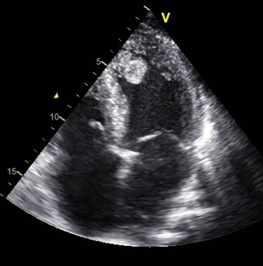
The main diagnostic tests for detecting thrombi in the left ventricle are transthoracic echocardiography (TTE) and cardiac magnetic resonance imaging (CMRI) with delayed gadolinium enhancement. TTE is the most used initial technique due to its availability and low cost: however, its sensitivity is limited (approximately 21-35%), although its specificity is high (95-98%). The use of intravenous contrast agents in TTE improves sensitivity (up to 64%) without losing specificity. Transthoracic echocardiography has been utilized for identifying left ventricular thrombi since the early 1980s. In recent years, the introduction of echocardiographic contrast agents has improved detections accuracy, particularly in patients with suboptimal acoustic windows. TTE remains the initial diagnostic modality of choice for evaluating left ventricular thrombus. However, its limitations such as difficulty imaging patients with poor acoustic windows, can lead to considerable interobserver variability, potentially compromising diagnostic reliability. Cardiac magnetic resonance offers a diagnostic edge over echocardiography by allowing both myocardial tissue characterization and dynamic imaging. With recent advancements in imaging sequences and the use of paramagnetic contrast agents to enhance blood pool visualization, late gadolinium enhancement CMR may offer superior sensitivity for detecting left ventricular thrombi.
Recent epidemiologic tests have provided that the incidence of left ventricular
thrombus, using optimal imaging modalities, can reach up to 15% in patients with ST segment elevation myocardial infarction and up to 25% in those with anterior myocardial infarction. Although a standard transthoracic echocardiogram is frequently used for initial screening, its low sensitivity in detecting left ventricular thrombus requires the use of contrast (when not contraindicated) and/or cardiac MRI when there is a high pretest probability.
Transesophageal echocardiography does not provide advantages for visualizing
the ventricular apex and is not recommended as a second-line method for
ventricular thrombi.
The first study that was able to compare the diagnostic accuracies of CMRI,
contrast TTE and noncontrast TTE was performed by Weinsaft et al. That
demonstrated that even with administration of echocardiographic contrast agents, CMRI was still considerably more accurate modality in terms of thrombus detection.
CMR with late gadolinium enhancement is the gold standard, with a sensitivity of 82-88% and specificity of 99-100%, as it allows differentiation of the thrombus (avascular without enhancement) from the surrounding myocardium. It is especially recommended when TTE (even with contrast) is not diagnostic or clinical suspicion persists. Cardiac computed tomography can incidentally detect thrombi, but it is not validated for this purpose
Precise detection of left ventricular thrombi is crucial, as it frequently guides the initiation of anticoagulation therapy to reduce the risk of embolic complications. While current guidelines suggest that starting anticoagulation may be reasonable in patients with strong suspicion of thrombus such as those with apical akinesis or dyskinesis even without visible thrombus, selecting the most appropriate imaging modality is essential to ensure timely and evidence-based therapeutic decisions.

Complications
The main complications of thrombi in the left ventricle are systemic embolic events, especially ischemic stroke and peripheral arterial embolisms. Embolization occurs because the thrombus can detach and migrate into systemic circulation, affecting organs such as the brain, kidneys, spleen, or extremities. The risk of embolization is particularly high in the first few weeks after an acute myocardial infarction and can reach up to 22% depending on the morphology and follow up of the thrombus.
The incidence of systemic embolic events in patients with left ventricular thrombi varies depending on the population and clinical context. In patients with acute myocardial infarction (AMI), the incidence of left ventricular thrombus is 3.5% to 7.1% after previous AMI when cardiac magnetic resonance imaging is used, and the incidence of systemic embolism (including stroke) in the presence of thrombus is between 7% and 16% in the first few years after the event, with an annualized risk of 3.7% compared to 0.8% in patients without left ventricular thrombus. Other relevant complications include major adverse cardiovascular events (MACE), which include death, reinfarction, and hospitalization for heart failure. The presence of left ventricular thrombus is associated with a significant increase in mortality and long-term adverse cardiovascular events. In addition, patients with persistent thrombus are at increased risk of bleeding, especially if they require prolonged anticoagulation.
The American Heart Association emphasizes that complete thrombus resolution is associated with lower mortality, while thrombus persistence, especially if mural and organized, carries a lower but not zero risk of embolization. The patient groups with the highest incidence of complications associated with thrombi in the left ventricle are mainly those with extensive acute myocardial infarction (AMI), especially anterior AMI, patients with ventricular aneurysm, and those with reduced left ventricular ejection fraction (LVEF). In addition, patients with dilated cardiomyopathy, either ischemic or non-ischemic, particularly those with severe systolic dysfunction, also have an elevated risk of embolic complications and major cardiovascular events.
In the context of non-ischemic cardiomyopathy, patients with dilated
cardiomyopathy show an even higher risk of systemic embolism compared to other non-ischemic etiologies and ischemic heart disease. The presence of mobile or protruding thrombi increases the risk of embolization, while thrombus persistence is associated with higher mortality and adverse events.
The American Heart Association points out that the combination of anterior AMI, low LVEF, ventricular aneurysm, and delayed reperfusion are factors that identify patients at higher risk of embolic complications and mortality associated with thrombi in the left ventricle.
The factors that increase the risk of thrombus formation in the left ventricle vary depending on the patient group, but they share pathophysiological mechanisms based on Virchow’s triad: ventricular dysfunction (stasis), endocardial damage, and inflammation/hypercoagulability. In patients with extensive acute myocardial infarction (AMI), especially anterior AMI, the highest risk factors are anterior location of the infarction, presence of ventricular aneurysm, left ventricular ejection fraction (LVEF) <30-40%, larger infarction size (elevated troponins), delayed reperfusion, and suboptimal coronary flow after intervention. The combination of reduced LVEF and segmental dysfunction (particularly apical) is the main predictor of thrombus and embolic complications or major cardiovascular events in all these groups. Systemic inflammation (elevated CRP) and the use of certain antithrombotic drugs may also contribute

Managment
Management of left Heart Thrombi (RHT)

The cornrstone of managment for intracardiac thrombus, particularly left ventricular thrombus, is therapeutic anticoagulantion. This strategy aims to reduce the risk of systemic embolism and promote trhombus resolution. Anticoagulation should be initiated promptly upon diagnosis, typically with intravenous unfractionated heparin, low molecular weight heparin, or a direct oral anticoagulant (DOAC). Transition to oral therapy with either warfarin or a DOAC is the recommended available evidence suggests that anticoagulation significantly lowers embolic risk and increases the likelihood of thrombus resolution compared to no or subtherapeutic treatment. In particular, a higher time in therapeutic range with warfarin is associated with superior outcomes and appears to outweigh the bleeding risks, even in the presence of concurrent antiplatelet therapy. The standard duration of anticoagulation is a minimum of three months.

Follow-up
cardiac imaging, ideally using the same modality employed at diagnosis, should be performed at that point to assess thrombus resolution. If the thrombus persists without notable change, anticoagulation should be continued with periodic reassessment. In cases where the thrombus has decreased in size or displays features consistent with chronicity and reduced embolic potential, the decision to continue therapy should be based on ongoing embolic risk, such as persistent left ventricular dysfunction, aneurysm formation, or spontaneous echocardiographic contrast. If both the thrombus and contributing risk factors have resolved, evidenced by normalization of systolic function and absence of additional indications for anticoagulation, discontinuation of therapy may be appropriate. For patients who develop LVT in the context of prior MI (≥3 months) or chronic ischemic cardiomyopathy, no randomized controlled data exist to guide treatment duration. Nonetheless, anticoagulation for a period of 3 to 6 months is generally recommended. Beyond that, extended or indefinite therapy should be considered on a case-by-case basis, incorporating individual thrombotic and bleeding risks, recovery of ventricular function, and patient preferences through shared decision-
making.

Management of Right Heart Thrombi (RHT)
Right heart thrombi (RHT) are rare but potentially life-threatening findings, often associated with pulmonary embolism (PE) and right ventricular dysfunction. The management of RHT remains a clinical challenge due to the lack of randomized controlled trials and standardized treatment guidelines. However, observational studies and registry data suggest that anticoagulation alone is often insufficient, especially in cases involving mobile or serpiginous thrombi with high embolic potential.
Initial management typically includes systemic anticoagulation with intravenous unfractionated heparin or low molecular weight heparin. This serves as a bridge to definitive therapy and may be appropriate in hemodynamically stable patients with non-mobile thrombi or contraindications to more aggressive interventions.
For patients with mobile RHT or hemodynamic compromise, reperfusion strategies are generally preferred. Systemic thrombolysis has demonstrated lower mortality rates compared to anticoagulation alone, but carries a notable risk of major bleeding, including intracranial hemorrhage. Surgical embolectomy is another option, particularly in patients with contraindications to thrombolysis or when thrombi are large, organized, or entangled in cardiac structures.
Catheter-directed therapies, including percutaneous aspiration thrombectomy (e.g., AngioVac, FlowTriever, AlphaVac), have gained attention as minimally invasive alternatives. These techniques allow for rapid thrombus removal with high success rates and a lower bleeding profile compared to systemic thrombolysis. Early outcomes are promising, although data remain limited and long-term efficacy has not been firmly established.
Ultimately, the choice of therapy should be guided by thrombus characteristics
(size, mobility, morphology), patient stability, comorbidities, bleeding risk, and institutional expertise. In general, mobile RHTs or those associated with acute PE warrant urgent intervention beyond anticoagulation alone. Multidisciplinary decision making often involving cardiology, critical care, interventional radiology, and cardiothoracic surgery is essential for optimizing outcomes.

Prevention
Intracardiac thrombus formation is a recognized complication in patients with heart failure and reduced ejection fraction, particularly in those with non-ischemic dilated
cardiomyopathy (DCM). Although left ventricular (LV) thrombi are more frequently documented, thrombi may also develop in the right heart chambers, especially in the presence of right-sided dysfunction, central venous catheters, cardiac devices, or systemic hypercoagulable states.
The use of antithrombotic therapy for primary prevention of thrombus formation in this population remains a subject of ongoing clinical judgment. In patients with DCM who are in sinus rhythm and without prior thromboembolic events, neither aspirin nor warfarin has consistently demonstrated clear benefit in preventing thrombus formation or reducing major adverse cardiovascular events. Therefore, routine prophylactic use of these agents is generally not recommended. However, individualized assessment is essential, especially when additional risk factors such as atrial fibrillation, prior embolic events, severely reduced ejection fraction, or left ventricular aneurysms are present.
In select subtypes of DCM that carry a higher inherent risk of intracardiac thrombus such as Takotsubo syndrome with apical ballooning, left ventricular
noncompaction, peripartum cardiomyopathy, eosinophilic myocarditis, and
infiltrative diseases like cardiac amyloidosis the use of oral anticoagulants (e.g.,
warfarin) or parenteral agents may be considered on a case-by-case basis. In
contrast, low-dose aspirin may offer some theoretical antiplatelet benefit, but its role in thrombus prevention remains less defined. Long-term anticoagulation may be appropriate for patients with persistent ventricular dysfunction or recurrent thromboembolic risk, provided the bleeding risk is acceptable.

Bibliografia link
 Mathevosian, S., & Ranade, M. (2022). Right Heart Clot-in-Transit: Endovascular
Therapies. Seminars in interventional radiology, 39(5), 515–522.
https://doi.org/10.1055/s-0042-1757942
 Sakellariou, X. M., Efstathopoulos, A., Stamatis, K. V., Nikas, D. N., & Kolettis, T.
M. (2020). Treatment of Mobile Right Heart Thrombi. European journal of case
reports in internal medicine, 7(12), 001918. https://doi.org/10.12890/2020_001918
 Patel, A. N., Amrutiya, R. J., Manvar, B. N., & Patel, A. (2022). A proposed
approach for the management of clot-in-transit. Cureus, 14(8).
 Lawrence LK Leung, M. D. (2025). Overview of hemostasis.
https://shorturl.at/hkxsH
 Levine, G. N., McEvoy, J. W., Fang, J. C., Ibeh, C., McCarthy, C. P., Misra, A.,
Shah, Z. I., Shenoy, C., Spinler, S. A., Vallurupalli, S., Lip, G. Y. H., on behalf of
the American Heart Association Council on Clinical Cardiology, Council on
Cardiovascular and Stroke Nursing, & and, S. C. (2022). Management of patients at
risk for and with left ventricular thrombus: A scientific statement from the american
heart association. Circulation, 146(15), e205–e223.
10.1161/CIR.0000000000001092
 Warren J Manning, M. D. (2024). Echocardiography in detection of cardiac and
aortic sources of systemic embolism. https://shorturl.at/mR7gN
 Watson, N. W., Weinberg, I., Dicks, A. B., Carroll, B. J., & Secemsky, E. A. (2024).
Clinical outcomes and predictors of advanced therapy for the management of right
heart thrombus. Circulation: Cardiovascular Interventions, 17(4), e013637.
10.1161/CIRCINTERVENTIONS.123.013637

 Wilson S Colucci, M. D., & Gregory YH Lip, MD, FRCPE, FESC, FACC. (2025).
Left ventricular thrombus. https://shorturl.at/Zf9oO
 Kleindorfer, D. O., Towfighi, A., Chaturvedi, S., Cockroft, K. M., Gutierrez, J.,
Lombardi-Hill, D., Kamel, H., Kernan, W. N., Kittner, S. J., Leira, E. C., Lennon,
O., Meschia, J. F., Nguyen, T. N., Pollak, P. M., Santangeli, P., Sharrief, A. Z.,
Smith, S. C., Jr, Turan, T. N., & Williams, L. S. (2021). 2021 guideline for the
prevention of stroke in patients with stroke and transient ischemic attack: A
guideline from the American heart association/American stroke association. Stroke;
a Journal of Cerebral Circulation, 52(7), e364–e467.
https://doi.org/10.1161/STR.0000000000000375
 Levine, G. N., McEvoy, J. W., Fang, J. C., Ibeh, C., McCarthy, C. P., Misra, A.,
Shah, Z. I., Shenoy, C., Spinler, S. A., Vallurupalli, S., Lip, G. Y. H., & American
Heart Association Council on Clinical Cardiology; Council on Cardiovascular and
Stroke Nursing; and Stroke Council. (2022). Management of patients at risk for and
with left ventricular thrombus: A scientific statement from the American Heart
Association. Circulation, 146(15), e205–e223.
https://doi.org/10.1161/CIR.0000000000001092
 Camaj, A., Fuster, V., Giustino, G., Bienstock, S. W., Sternheim, D., Mehran, R.,
Dangas, G. D., Kini, A., Sharma, S. K., Halperin, J., Dweck, M. R., & Goldman, M.
E. (2022). Left ventricular thrombus following acute myocardial infarction: JACC
state-of-the-art review. Journal of the American College of Cardiology, 79(10),
1010–1022. https://doi.org/10.1016/j.jacc.2022.01.011

 Sharma, N. D., McCullough, P. A., Philbin, E. F., & Weaver, W. D. (2000). Left
ventricular thrombus and subsequent thromboembolism in patients with severe
systolic dysfunction. Chest, 117(2), 314–320.
https://doi.org/10.1378/chest.117.2.314
 Ram, P., Shah, M., Sirinvaravong, N., Lo, K. B., Patil, S., Patel, B., Tripathi, B.,
Garg, L., & Figueredo, V. (2018). Left ventricular thrombosis in acute anterior
myocardial infarction: Evaluation of hospital mortality, thromboembolism, and
bleeding. Clinical Cardiology, 41(10), 1289–1296. https://doi.org/10.1002/clc.23039
 Albaeni, A., Chatila, K., Beydoun, H. A., Beydoun, M. A., Morsy, M., & Khalife,
W. I. (2020). In-hospital left ventricular thrombus following ST-elevation
myocardial infarction. International Journal of Cardiology, 299, 1–6.
https://doi.org/10.1016/j.ijcard.2019.07.070

Bacterial Endocarditis Prophylaxis

Dr. Pakeeza Saif
King Edward Medical College, Lahore, Pakistan


Amin H. Karim MD

Houston Methodist Academic Institute
and Weill Medical College of


Dear Dentist: My Murmur Doesn’t Need Meds Anymore!

Infective endocarditis (IE) is a serious infection of the heart’s inner lining, affecting 3 to 10 people per 100,000 annually. It carries a significant risk, with mortality reaching up to 30% within the first 30 days 1 . Staphylococcus aureus was the most frequently identified pathogen, accounting for 31% of cases. The mitral valve was the most commonly affected, involved in forty-one percent of infections, while the aortic valve was affected in thirty-eight percent of cases 2. The diagnosis of IE is primarily clinical and is based on the modified Duke criteria, which include a combination of major and minor clinical, microbiological, and echocardiographic findings.

Guntheroth et al. observed that bacteremia was present in 40% of 2,403 cases following tooth extraction, 38% of individuals during routine mastication, and 11% of those with oral sepsis in the absence of any dental intervention 3. This issue has long concerned both dentists and cardiologists, driven in part by a preference for commission bias—favoring action over inaction—when considering prophylactic antibiotic use.

American Heart Association revised the guidelines on infective endocarditis prophylaxis in 2007 (full guidelines available at http://circ.ahajournals.org ) to promote the judicious use of antibiotics, particularly in clinical scenarios where the anticipated benefits are outweighed by the risks, such as the emergence of antibiotic resistance and the potential for adverse drug reactions. The present revised document was not based on the results of a single study but rather on the collective body of evidence published in numerous studies over the past two decades. The following points were used as a rationale by AHA for updating the guideline4.

  1. IE is much more likely to result from frequent exposure to random bacteremia associated with daily activities such as chewing food, tooth brushing, flossing, use of toothpicks, use of water irrigation devices, and other activities than from bacteremia caused by a dental, gastrointestinal (GI) tract or genitourinary (GU) tract procedure.
  2. Prophylaxis prevents only an exceedingly small number of cases of IE, if any, in individuals who undergo a dental, GI tract, or GU tract procedure.
  3. The risk of antibiotic-associated adverse events exceeds the benefit, if any, from prophylactic antibiotic therapy except in very high-risk situations.
  4. Maintenance of optimal oral health and hygiene may reduce the incidence of bacteremia from daily activities and thus the risk of IE and is more important than the use of prophylactic antibiotics for dental procedures 4.

Several studies have demonstrated that the lifetime risk of infective endocarditis (IE) varies significantly depending on the underlying cardiac condition. In the general population without known heart disease, the risk is approximately 5 cases per 100,000 patient-years. Patients with rheumatic heart disease (RHD) face a substantially higher risk, ranging from 380 to 440 cases per 100,000 patient-years, which is comparable to the risk observed in individuals with mechanical or bioprosthetic heart valves (308 to 383 cases per 100,000 patient-years)5 .

The greatest risks are observed in the following groups:

  • 630 cases per 100,000 patient-years following cardiac valve replacement for native valve IE
  • 740 cases per 100,000 patient-years in patients with a history of previous IE
  • 2,160 cases per 100,000 patient-years in patients undergoing prosthetic valve replacement due to prosthetic valve endocarditis

These variations in risk highlight the importance of tailoring preventive measures to individual patient profiles.

Further research indicates that even with perfect effectiveness, antibiotic prophylaxis would prevent only a negligible number of infective endocarditis cases—given that the estimated absolute risk after a dental procedure is about 1 in 1.1 million for mitral valve prolapse, 1 in 475 000 for congenital heart disease, 1 in 142 000 for rheumatic heart disease, 1 in 114 000 for prosthetic heart valves, and 1 in 95 000 for those with a history of endocarditis6 7 .

Cardiac Conditions Associated with the Highest Risk of Adverse Outcome from Endocarditis for Which Prophylaxis Is Reasonable

  • Prosthetic cardiac valve or prosthetic material used for cardiac valve repair
  • Previous Infective Endocarditis
  • Cardiac transplantation recipients who develop cardiac valvulopathy
  • Congenital heart disease (CHD)
  • Unrepaired cyanotic CHD, including palliative shunts and conduits
  • Completely repaired congenital heart defect with prosthetic material or device, whether placed by surgery or by catheter intervention, during the first 6 months after the procedure
  • Repaired CHD with residual defects at the site or adjacent to the site of a prosthetic patch or prosthetic device.

Guidelines also clearly stated that antibiotic prophylaxis is no longer recommended for any other form of congenital heart disease which explicitly includesheart murmurs, valvular regurgitation, or stenosis without prosthetic material or prior endocarditis 4.

Preventing Infective Endocarditis: Procedures Requiring Antimicrobial Prophylaxis

High-Risk Procedures Requiring Antibiotic Prophylaxis

  • Dental procedures that involve manipulation of gingival tissue or the periapical region of teeth or perforation of the oral mucosa
  • Respiratory tract procedure with incision and biopsy such as tonsillectomy and adenoidectomy
  • Gastrointestinal or genitourinary procedures in setting of active infection
  • Surgery on infected skin, skin structure, or musculoskeletal tissue

Low-Risk Procedures Not Requiring Antibiotic Prophylaxis

  • Gastrointestinal or Genitourinary procedure in the absence of infection
  • Most Vaginal deliveries or Caesarian deliveries
  • Left atrial appendage occlusion device placement (e.g., Watchman) in the absence of infection — associated with a very low incidence of infective endocarditis, with long-term studies showing no device-related infections over extended follow-up. A single center, 14 year study of 181 patients found no device-related infections over more than 500 patient years of follow up8 .
  • Stable cardiac implantable electronic devices (CIEDs) such as pacemakers and ICDs — antibiotic prophylaxis is not recommended for dental or mucosal procedures solely due to the presence of a CIED in the absence of other high-risk cardiac conditions9 .
  • Atrial septal defect (ASD) closure devices beyond 6 months post-implantation — prophylaxis is not indicated once the device is fully endothelialized and no residual shunt remains10 .

Infective Endocarditis Prophylaxis: Antibiotic Recommendations

  • First line: 2 g amoxicillin orally (or 50 mg/kg kids) 30–60 minutes before the procedure.
  • If allergic to penicillin, 600 mg clindamycin orally (or 20 mg/kg kids).
  • Alternatives include 500 mg azithromycin or clarithromycin orally (15 mg/kg kids)
  • If you can’t take pills, get 2 g ampicillin IM/IV (or 50 mg/kg kids)

Considering rising antimicrobial resistance and the potential for Clostridioides difficile infection linked to antibiotic use, it is advised against relying on the outdated “better safe than sorry” approach to prophylactic antibiotic use, as it may cause more harm than benefit to patients.

References

1. Mostaghim AS, Lo HYA, Khardori N. A retrospective epidemiologic study to define risk factors, microbiology, and clinical outcomes of infective endocarditis in a large tertiary-care teaching hospital. SAGE Open Med. 2017;5. doi:10.1177/2050312117741772

2. Murdoch DR. Clinical Presentation, Etiology, and Outcome of Infective Endocarditis in the 21st Century. Arch Intern Med. 2009;169(5):463. doi:10.1001/archinternmed.2008.603

3. Guntheroth WG. How important are dental procedures as a cause of infective endocarditis? Am J Cardiol. 1984;54(7):797-801. doi:10.1016/S0002-9149(84)80211-8

4. Wilson W, Taubert KA, Gewitz M, et al. Prevention of Infective Endocarditis. Circulation. 2007;116(15):1736-1754. doi:10.1161/CIRCULATIONAHA.106.183095

5. Steckelberg JM; WWR. Risk factors for infective endocarditis. Infectious disease clinics of North America. 1993;7(1):9-19.

6. Pallasch TJ, Wahl MJ. Focal infection: new age or ancient history? Endod Topics. 2003;4(1):32-45. doi:10.1034/j.1601-1546.2003.00002.x

7. Pallasch TJ. Antibiotic prophylaxis: problems in paradise. Dent Clin North Am. 2003;47(4):665-679. doi:10.1016/S0011-8532(03)00037-5

8. Ward RC, McGill T, Adel F, et al. Infection Rate and Outcomes of Watchman Devices: Results from a Single-Center 14-Year Experience. Biomed Hub. 2021;6(2):59-62. doi:10.1159/000516400

9. Canpolat U. Tailored antibiotic prophylaxis in patients undergoing CIED implantation: One size does not fit all the principle. Pacing and Clinical Electrophysiology. 2019;42(4):483-483. doi:10.1111/pace.13624

10. Tanabe Y, Sato Y, Izumo M, et al. Endothelialization of an Amplatzer Septal Occluder Device 6 Months Post Implantation: Is This Enough Time? An In Vivo Angioscopic Assessment. Journal of Invasive Cardiology. 2019;31(2). doi:10.25270/jic/18.00206

How Low is Low?

RISKS OF VERY LOW LDL CHOLESEROL LEVEL

By Laura Edith Chavez Salas
Universidad De Durango, Campus Zacatecas, Mexico

Amin H. Karim, MD
Houston Methodist Academic Institute

Low-density lipoprotein (LDL) represents a category of lipoprotein particles responsible for the transport of cholesterol and various lipids within the bloodstream. Often referred to as the “bad” cholesterol, it serves vital purposes. LDL particles serve as the primary carriers of cholesterol to peripheral tissues and consist of cholesteryl esters and triglycerides encased in a phospholipid shell, free cholesterol, and a single molecule of apolipoprotein B-100. Increased levels of LDL are directly associated with the onset of atherosclerotic cardiovascular disease (ASCVD), as LDL particles can penetrate the arterial wall, become retained and altered (for instance, oxidized), and facilitate the development of foam cells and atherosclerotic plaques. (1-4)

LDL exhibits heterogeneity, with subclasses that vary in size and density; smaller, denser LDL particles are deemed more atherogenic compared to their larger, more buoyant counterparts. (3, 5, 6) The cholesterol content within LDL particles is quantified as LDL cholesterol (LDL-C), which serves as a conventional marker for evaluating and managing cardiovascular risk. (7) Nevertheless, the quantity of LDL particles (LDL-P) and the concentration of apolipoprotein B (apoB) may offer further risk stratification, particularly in individuals with metabolic syndrome or diabetes, as discrepancies between LDL-C and LDL-P can arise. (8) Evaluation of ASCVD risk can be evaluated by assessing both LDL-C and LDL-P, asserting that the reduction of LDL—primarily through the use of statins and other lipid-lowering treatments—leads to a decrease in cardiovascular events. (4)

DANGERS OF VERY LOW LDL

However, an LDL-lowering regimen can lead to ultra-low-density lipoprotein cholesterol (LDL-C) levels. These are typically defined as <40–50 mg/dL, and especially <30 mg/dL. These levels are generally well tolerated and associated with a reduced risk of atherosclerotic cardiovascular disease (ASCVD), but several potential dangers have been identified. (9) Mechanistically, very low LDL-C may impair endothelial integrity and platelet function. This could potentially increase bleeding risk, especially for intracranial and gastrointestinal hemorrhage. (10, 11)

The most observed danger of ultra-low LDL is a possible hemorrhagic stroke and other bleeding events, particularly at LDL-C levels below 40 mg/dL, as supported by mechanistic and clinical data. Observational studies and meta-analyses have also reported a U-shaped relationship between LDL-C and all-cause mortality, with both very low (<50 mg/dL) and high (≥130 mg/dL) LDL-C levels associated with increased mortality in certain populations, such as those with coronary artery disease. (15)

There is also some evidence suggesting a potential association between ultra-low LDL-C and increased risk of new-onset diabetes mellitus, particularly with statin therapy. Leading to more complications, there is a possible link to cataract formation and glaucoma, though causality remains unproven and the absolute risk is low. (11, 14)

The main dangers of ultra-low LDL-C are a possible increased risk of hemorrhagic stroke, new-onset diabetes, and, less consistently, all-cause mortality in specific populations. However, for most high-risk patients, the cardiovascular benefits of aggressive LDL-C lowering outweigh these potential risks. (9-14)

HIGH-RISK PATIENTS

Patients at highest risk for complications associated with very low levels of low-density lipoprotein cholesterol (LDL-C) are:

• Individuals with a prior history of hemorrhagic stroke:

The American Stroke Association notes that the risk of hemorrhagic stroke with statin therapy is small and nonsignificant in those without prior cerebrovascular disease, but patients with a history of hemorrhagic stroke may be at increased risk, and lipid lowering in this group requires individualized consideration and further study. (16)

• Patients with poorly controlled hypertension and very low LDL-C:

There is literature that indicates that the combination of very low LDL-C (especially ≤40 mg/dL) and uncontrolled hypertension substantially increases the risk of both ischemic and hemorrhagic stroke. Although this risk is particularly more prevalent in East Asian populations, it is relevant globally. (17)

• Women with LDL-C <70 mg/dL:

There is evidence from long-term cohort studies in women that has shown that LDL-C <70 mg/dL is associated with a more than twofold increased risk of hemorrhagic stroke compared to LDL-C 100–129.9 mg/dL, independent of other risk factors. Meaning that women with no other risk factors have more risk than males with no other risk factors. (18)

• Patients on intensive statin therapy or with other risk factors for diabetes: Statin therapy, especially at high intensity, is associated with a modestly increased risk of new-onset diabetes. Particularly in those with predisposing factors such as metabolic syndrome or impaired fasting glucose. (11)

Risks associated with having ultra-low LDL-C are more prevalent in populations most at risk, which are those with prior hemorrhagic stroke, poorly controlled hypertension, women, and individuals with multiple vascular risk factors or on intensive lipid-lowering therapy. 

REFERENCES

  1. Orlova, E V et al. “Three-dimensional structure of low density lipoproteins by electron cryomicroscopy.” Proceedings of the National Academy of Sciences of the United States of America vol. 96,15 (1999): 8420-5. doi:10.1073/pnas.96.15.8420
  2. Rhainds, D, and L Brissette. “Low density lipoprotein uptake: holoparticle and cholesteryl ester selective uptake.” The international journal of biochemistry & cell biology vol. 31,9 (1999): 915-31. doi:10.1016/s1357-2725(99)00046-1
  3. Qiao, Ya-Nan et al. “Low-density lipoprotein particles in atherosclerosis.” Frontiers in physiology vol. 13 931931. 30 Aug. 2022, doi:10.3389/fphys.2022.931931
  4. Maurya, Rupesh et al. “Low density lipoprotein receptor endocytosis in cardiovascular disease and the factors affecting LDL levels.” Progress in molecular biology and translational science vol. 194 (2023): 333-345. doi:10.1016/bs.pmbts.2022.09.010
  5. Ivanova, Ekaterina A et al. “Small Dense Low-Density Lipoprotein as Biomarker for Atherosclerotic Diseases.” Oxidative medicine and cellular longevity vol. 2017 (2017): 1273042. doi:10.1155/2017/1273042
  6. Packard, C et al. “The role of small, dense low density lipoprotein (LDL): a new look.” International journal of cardiology vol. 74 Suppl 1 (2000): S17-22. doi:10.1016/s0167-5273(99)00107-2
  7. Jialal, I, and A T Remaley. “Measurement of low-density lipoprotein cholesterol in assessment and management of cardiovascular disease risk.” Clinical pharmacology and therapeutics vol. 96,1 (2014): 20-2. doi:10.1038/clpt.2014.69
  8. Galimberti, Federica et al. “Apolipoprotein B compared with low-density lipoprotein cholesterol in the atherosclerotic cardiovascular diseases risk assessment.” Pharmacological research vol. 195 (2023): 106873. doi:10.1016/j.phrs.2023.106873
  9. Karagiannis, Angelos D et al. “How low is safe? The frontier of very low (<30 mg/dL) LDL cholesterol.” European heart journal vol. 42,22 (2021): 2154-2169. doi:10.1093/eurheartj/ehaa1080
  10. Siniscalchi, Carmine et al. “Low LDL-Cholesterol and Hemorrhagic Risk: Mechanistic Insights and Clinical Perspectives.” International journal of molecular sciences vol. 26,12 5612. 11 Jun. 2025, doi:10.3390/ijms26125612
  11. Cure, Erkan, and Medine Cumhur Cure. “Emerging risks of lipid-lowering therapy and low LDL levels: implications for eye, brain, and new-onset diabetes.” Lipids in health and disease vol. 24,1 185. 21 May. 2025, doi:10.1186/s12944-025-02606-6
  12. Olsson, A G et al. “Can LDL cholesterol be too low? Possible risks of extremely low levels.” Journal of internal medicine vol. 281,6 (2017): 534-553. doi:10.1111/joim.12614
  13. Rong, Shuang et al. “Association of Low-Density Lipoprotein Cholesterol Levels with More than 20-Year Risk of Cardiovascular and All-Cause Mortality in the General Population.” Journal of the American Heart Association vol. 11,15 (2022): e023690. doi:10.1161/JAHA.121.023690
  14. Faselis, Charles et al. “Is very low LDL-C harmful?.” Current pharmaceutical design vol. 24,31 (2018): 3658-3664. doi:10.2174/1381612824666181008110643
  15. Scudeler, Thiago Luis et al. “Association between low-density lipoprotein cholesterol levels and all-cause mortality in patients with coronary artery disease: a real-world analysis using data from an international network.” Scientific reports vol. 14,1 29201. 25 Nov. 2024, doi:10.1038/s41598-024-80578-w
  16. Goldstein, Larry B., et al. “Aggressive LDL-C Lowering and the Brain: Impact on Risk for Dementia and Hemorrhagic Stroke: A Scientific Statement From the American Heart Association.” Arteriosclerosis Thrombosis and Vascular Biology, vol. 43, no. 10, Sept. 2023, https://doi.org/10.1161/atv.0000000000000164.
  17. Wu, Zhijun et al. “The risk of ischemic stroke and hemorrhagic stroke in Chinese adults with low-density lipoprotein cholesterol concentrations < 70 mg/dL.” BMC medicine vol. 19,1 142. 16 Jun. 2021, doi:10.1186/s12916-021-02014-4
  18. Rist, Pamela M et al. “Lipid levels and the risk of hemorrhagic stroke among women.” Neurology vol. 92,19 (2019): e2286-e2294. doi:10.1212/WNL.0000000000007454

Dupuytren’s Contracture

Authors:
Tejaswini Potlabathini,
Elaine Tran,
PA Student, University of Texas Medical Branch, Galveston, Tx.
Amin H. Karim MD FRCP FACC

Patient Overview

A 72-year-old female with a past medical history of hypertension, hyperlipidemia, hypothyroidism, and prediabetes presented to the cardiology clinic for a routine follow-up. On examination, she was found to have a firm, nodular thickening of tissue on one palm over the third metacarpal area, associated with nearby skin puckering, prompting a presumptive diagnosis of Dupuytren’s contracture. A close up of a hand

AI-generated content may be incorrect.

Description

Dupuytren’s contracture is a progressive fibroproliferative disorder of the palmar fascia that leads to flexion contractures of the digits. It affects approximately 0.6% to 31.6% of the general population, with prevalence increasing with age. While its exact cause remains unclear, it is believed to have a multifactorial etiology involving both genetic and environmental factors. Genetically, it follows an autosomal dominant inheritance pattern more commonly in individuals of Northern European descent. The condition predominantly affects men but can also occur in women, often with a later onset and milder progression. Dupuytren’s contracture is associated with other fibrotic disorders, including plantar fibromatosis (Ledderhose disease) and dorsal hand nodules (Garrod pads). Several risk factors have been identified, including diabetes mellitus, smoking, and chronic alcohol consumption, all of which may contribute to microvascular ischemia and tissue damage. Additionally, the condition has been linked to seizure disorders and chronic liver disease, particularly in patients with cirrhosis. Occupational risk factors, such as repetitive manual labor and prolonged exposure to hand-transmitted vibration, may also accelerate disease progression.

Etiology

Fibroblasts, which are mesenchymal cells responsible for tissue maintenance, play a crucial role in the pathology of Dupuytren’s contracture. The disease progresses through three stages: proliferative, involutional, and residual. In the proliferative stage, fibroblasts are stimulated and differentiate into mature myofibroblasts under the influence of transforming growth factor-beta (TGF-β) and mechanical stress from associated risk factors. As the disease advances to the involutional stage, nodules begin to form, producing an extracellular matrix (ECM) rich in type III collagen. In the residual stage, fibrotic tissue stabilizes as the ratio of type I to type III collagen increases, leading to collagen cross-linking. This results in the formation of fibrous cords, which cause progressive digital flexion contractures. Additionally, the presence of CD3-positive lymphocytes and the expression of major histocompatibility complex (MHC) class II proteins suggest a possible role for a T-cell-mediated autoimmune response in the disorder. In summary, Dupuytren’s contracture arises from fibroblastic proliferation and disorganized collagen deposition, ultimately leading to palmar fascial thickening and contracture formation.

A diagram of a hand with different stages of skin disease

AI-generated content may be incorrect.

Image Source: https://www.sciencedirect.com/science/article/pii/S0363502323000709

Clinical Presentation

Patients with Dupuytren’s contracture may present with painful or painless lumps in the palm, along with restricted finger mobility and decreased grip strength. As the nodules thicken and fibrous cords form, patients may experience difficulty straightening or spreading their fingers due to flexion contractures. The fourth and fifth digits are most commonly affected, with contractures typically involving the metacarpophalangeal (MCP) joint first, followed by the proximal interphalangeal (PIP) joint, and less frequently, the distal interphalangeal (DIP) joint.

Physical Examination 

On physical examination, the Hueston Tabletop Test can be performed. Patients are asked to place their palm flat on a table, and failure to do so indicates a positive test. If contractures are present, the angles at the MCP and PIP joints should be measured to assess disease severity and progression. Grading of Dupuytren’s contracture is as follows: Grade 1 presents as a thickened nodule and band in the palmar aponeurosis, which may progress to skin tethering, puckering, or pitting. Grade 2 presents as a peritendinous band, leading to limited extension of the affected finger. Grade 3 presents as a significant flexion contracture.

A close-up of hands

AI-generated content may be incorrect.

Treatment

Although there is no definitive cure for Dupuytren’s contracture, symptoms can be managed through nonsurgical and surgical options. Nonsurgical options include corticosteroid injections, collagenase clostridium histolyticum injections, and needle aponeurotomy. Corticosteroids alleviates symptoms by reducing inflammation while targeted collagenase injections can target and enzymatically degrade the collagen. In needle aponeurotomy, a fine needle is used to precisely cut through the fibrous cord. While this minimally invasive procedure does not remove the cord, a break in the cord allows for improved finger motion. Although more invasive and extensive, a partial palmar fasciectomy removes the abnormal tissue fibrous tissue and cords. Postoperative care includes splinting, wound care, and physical therapy. Emerging therapies include use of anti-tumor necrosis factors such as adalimumab injections to slow disease progression by targeting inflammatory pathways.  


References: 

  1. American Academy of Orthopaedic Surgeons. Dupuytren’s disease. OrthoInfo. Updated March 2023. Accessed January 25, 2025. https://orthoinfo.aaos.org/en/diseases–conditions/dupuytrens-disease/
  2. Dupuytren’s contracture. UpToDate. Updated January 15, 2025. Accessed January 25, 2025. https://www.uptodate.com/contents/dupuytrens-contracture
  3. Hindocha S, Stanley JK, Watson S, Bayat A. Revised Tubiana’s staging system for assessment of disease severity in Dupuytren’s disease—preliminary clinical findings. EBioMedicine. 2018;36:86-90. doi:10.1016/j.ebiom.2018.06.022.
  4. National Library of Medicine. Dupuytren contracture. MedlinePlus Genetics. Updated June 17, 2023. Accessed January 25, 2025. https://medlineplus.gov/genetics/condition/dupuytren-contracture/
  5. Zarb RM, Graf AR, Talhelm JE, et al. Dupuytren’s contracture recurrence and treatment following collagenase Clostridium histolyticum injection: a longitudinal assessment in a veteran population. Mil Med. 2023;188(9-10):e2975-e2981. doi:10.1093/milmed/usad075.

Adenocarcinoma and Pulmonary Tumor Thrombotic Microangiopathy.

By Emin Gayibov
Amin H. Karim MD

A Rapid Review of Adenocarcinoma and Pulmonary Tumor Thrombotic Microangiopathy: A Deadly Duo

Emin Gayibov • Amin H. Karim

Published: January 03, 2025 DOI: 10.7759/cureus.76842 

Peer-Reviewed

Cite this article as: Gayibov E, Karim A H (January 03, 2025) A Rapid Review of Adenocarcinoma and Pulmonary Tumor Thrombotic Microangiopathy: A Deadly Duo. Cureus 17(1): e76842. doi:10.7759/cureus.76842

Abstract

Pulmonary tumor thrombotic microangiopathy (PTTM) is a rare paraneoplastic syndrome associated with various adenocarcinomas, most commonly gastric adenocarcinoma. This condition can progressively worsen pulmonary arterial hypertension, leading to acute or subacute pulmonary heart failure and respiratory insufficiency. This paper examines the pathogenesis, clinical presentation, diagnosis, treatment, and prognosis of PTTM. Given PTTM’s poor prognosis, we emphasize treatment strategies. PTTM in adenocarcinoma patients can mimic other pulmonary diseases, causing diagnostic delays. Current PTTM treatment strategies primarily focus on managing the underlying malignancy and addressing thrombotic complications. Anti-angiogenic therapy with bevacizumab and the platelet-derived growth factor receptor antagonist imatinib have shown promise in multiple cases. Further research is needed to develop more effective and targeted therapies for this challenging condition. The precise mechanisms underlying this association remain to be fully elucidated.

Introduction & Background

Paraneoplastic syndromes constitute a diverse group of clinical disorders associated with malignant diseases that can significantly impact patient morbidity and mortality. They are not directly attributable to the physical effects of the primary or metastatic tumor [1]. In these syndromes, malignant cells do not directly cause symptoms related to metastasis; rather, they induce the generation of autoantibodies, cytokines, hormones, or peptides that exert effects on multiple organ systems [2]. It is estimated that paraneoplastic syndromes affect up to 8% of patients with cancer [3]. Table 1 provides a concise overview of commonly encountered paraneoplastic syndromes [4-23].

Organ systemParaneoplastic syndromeMost associated cancer typeClinical signs
NeurologicalParaneoplastic encephalitisSCLC, testicular cancerCognitive dysfunction, seizures, personality changes, hallucinations, autonomic dysfunction
Subacute cerebellar degenerationBreast, ovarian, SCLC, Hodgkin lymphomaAtaxia, dysarthria, dizziness, diplopia, nausea, vomiting
Opsoclonus-myoclonus syndromeNeuroblastoma (children), SCLC (adults)Rapid eye movements, body jerks, ataxia, hypotonia
Myasthenia gravisThymomaMuscle weakness, ptosis, diplopia, diaphragmatic weakness
Lambert-Eaton myasthenic syndromeSCLCProximal muscle weakness, autonomic dysfunction, diminished reflexes
Autonomic neuropathySCLC, thymomaDry mouth, altered pupillary reflexes, orthostatic hypotension, GI dysfunction
Subacute sensory neuropathySCLCParesthesia, neuropathic pain, diminished sensation
EndocrineCushing syndromeSCLC, pancreatic cancerHypertension, centripetal obesity, hypokalemia, edema
SIADHSCLCHyponatremia, lethargy, confusion, seizures
HypercalcemiaLung, renal cell, multiple myelomaLethargy, nausea, bradycardia, short QT interval on ECG
Non-islet tumor hypoglycemiaFibrosarcoma, hepatocellular carcinomaHypoglycemia, confusion, seizures
Carcinoid syndromeBronchial carcinoid, pancreatic carcinomaFlushing, diarrhea, bronchospasm
HyperaldosteronismAdrenal adenoma, non-Hodgkin lymphomaHypertension, hypokalemia
RheumatologicalParaneoplastic polyarthritisVarious malignanciesMigratory, asymmetric arthritis
Polymyalgia rheumaticaMyelodysplastic syndromePain and stiffness in shoulders, neck, and hips
Hypertrophic osteoarthropathyLung cancerDigital clubbing, joint swelling, periostitis
Multicentric reticulohistiocytosisVarious malignanciesPapules, nodules, destructive polyarthritis
HematologicalPolycythemiaRenal cell carcinoma, cerebellar hemangiomaIncreased hemoglobin, pallor, fatigue
Trousseau syndrome (migratory thrombophlebitis)Pancreatic, Bronchogenic carcinomaPainful migratory thrombophlebitis
DermatologicalAcanthosis nigricansGastric adenocarcinomaHyperpigmented, velvety plaques in axilla, neck
Paraneoplastic pemphigusB-cell lymphoproliferative disordersBlisters, mucosal erosions
Sweet syndromeHematological malignanciesPainful, erythematous plaques with fever
DermatomyositisOvarian, lung, pancreatic cancerHeliotrope rash, Gottron papules, proximal muscle weakness
RenalNephrotic syndromeVarious tumorsProteinuria, edema, fluid overload
Electrolyte imbalancesSCLC, thymomaHyponatremia, hyperphosphatemia, acid-base disturbances
PulmonaryPulmonary tumor thrombotic microangiopathyVarious adenocarcinomasMicrovascular thrombosis, endothelial injury, and severe pulmonary arterial hypertension
Lymphangitic carcinomatosisVarious adenocarcinomasProgressive dyspnea, cough, and hypoxemia
Pulmonary alveolar proteinosisLeukemia, lymphomaProgressive respiratory insufficiency
Table 1: A concise overview of paraneoplastic syndromes including pulmonary tumor thrombotic microangiopathy

Source: [4-23]

SCLC, small cell lung carcinoma; SIADH, syndrome of inappropriate antidiuretic hormone secretion

Pulmonary tumor thrombotic microangiopathy (PTTM) is a rare and often underrecognized paraneoplastic syndrome associated with various adenocarcinomas, often linked to gastric adenocarcinoma [24]. Although less frequently observed, breast, lung, and urothelial malignancies have also been linked to PTTM [25]. Post-mortem examinations of carcinoma patients indicate a PTTM prevalence ranging from 1.4% to 3.3% [24,26]. This condition can lead to a progressive deterioration of pulmonary arterial hypertension (PAH), culminating in acute or subacute pulmonary heart failure and respiratory insufficiency. Hence, early recognition and treatment are crucial to prevent irreversible lung damage and complications. First described by von Herbay et al. in 1990 [26], PTTM is characterized by the presence of numerous tumor emboli within the pulmonary microvasculature, leading to microvascular thrombosis and endothelial injury. Later studies have highlighted the critical role of tumor-derived factors in driving hypercoagulability and angiogenesis, as explained in the pathogenesis section.

In this paper, we will examine the pathogenesis, clinical presentation, diagnosis, treatment, and prognosis of PTTM in patients with adenocarcinoma of various origins. Given the poor prognosis associated with PTTM, we will place particular emphasis on treatment strategies. We will discuss the approaches taken in different cases in recent years to inform current best practices, including their limitations and potential avenues for improvement. This rapid review will delve into the current understanding of this rare paraneoplastic syndrome and its potential clinical implications. By reviewing the available literature, we aim to shed light on the pathophysiology, clinical manifestations, diagnostic challenges, and therapeutic options for PTTM.

Review

Methodology

All the studies referred for this rapid review have been searched via the National Library of Medicine (NIH), PubMed, with the free-text keywords “pulmonary tumor thrombotic microangiopathy (PTTM)”, “PTTM AND adenocarcinoma”, “pathogenesis AND PTTM”, “clinical presentation AND PTTM”, “diagnosis AND PTTM”, “treatment AND PTTM”, and “prognosis AND PTTM”. A visual tool Connected Papers [27] and reference manager software Mendeley [28] served for proper citation and access to the studies regarding the abovementioned aspects of PTTM. For this rapid review, we did not specify the time range of studies referred due to insufficiency in number. Our inclusion criteria comprised type I tumor embolism, which is the “classic” or “true” type originating from a distant primary tumor via hematologic seeding with no invasion into vessel walls. Among type I tumor embolism studies, we included ones that specifically focused on the PTTM, either antemortem or postmortem, as a confirmed diagnosis. We disregarded the studies involving type II tumor embolism, which results from a tumor growing into the pulmonary arteries with invasion into vessel walls. The search strategy is summarized in Table 2.

ItemsSpecification
Date of search1 October 2024 to 17 November 2024
Databases and other sources searchedNational Library of Medicine, PubMed
Search terms used“pulmonary tumor thrombotic microangiopathy (PTTM)”, “PTTM AND adenocarcinoma”, “pathogenesis AND PTTM”, “clinical presentation AND PTTM”, “diagnosis AND PTTM”, “treatment AND PTTM”, and “prognosis AND PTTM”
TimeframeNo specific timeframe
Inclusion and exclusion criteriaCase reports and reviews were included if they specifically focused on PTTM as type I tumor embolism, the “classic” or “true” type originating from a distant primary tumor via hematologic seeding without vessel wall invasion. Studies involving type II tumor embolism, resulting from a tumor growing into the pulmonary arteries with vessel wall invasion, and mixed embolism were excluded
Selection processA total of 164 studies were initially identified, of which 55 were ultimately included Author E.G. conducted the literature selection. A.K. supervised the search strategy.
Any additional considerationsConnected Papers [27] and Mendeley [28] served for proper citation and access to the studies
Table 2: Summary of the search strategy for this rapid review

Pathogenesis of pulmonary tumor thrombotic microangiopathy

Associated with mucin-secreting adenocarcinomas, PTTM is a specific type of pulmonary tumor embolism. It is classified within type I tumor embolism, the “classic” or “true” type originating from a distant primary tumor via hematologic seeding without vessel wall invasion [29,30]. In contrast, type II tumor embolism arises from a tumor growing into the pulmonary arteries with invasion into vessel walls [31]. In terms of histopathology, PTTM is characterized by fibrocellular intimal proliferation of small and medium pulmonary arteries and arterioles with the presence of tumor emboli [26,32]. This distinctive feature can be observed using various histological staining techniques, such as Verhoeff-Van Gieson and alpha-smooth muscle actin immunohistochemistry [33,34]. Fibrocellular intimal proliferation involves the growth of cells, primarily smooth muscle cells and fibroblasts, and extracellular matrix within the intima of small pulmonary arteries and arterioles. This leads to narrowing of the vessel lumen. Reactive fibrointimal thickening and occlusion were also revealed in the pulmonary lymphatic vessels and veins in a particular PTTM case [35]. It has also been reported that PTTM is frequently associated with lymphangiosis carcinomatosa, a serious condition that occurs when cancer cells spread to the lymph vessels, causing inflammation and blockage and becoming a predisposing factor to PTTM [26,36]. In PTTM, multiple microscopic tumor cells become stuck to the inner lining of small pulmonary blood vessels. This triggers an inflammatory response, causing the vessel walls to thicken and narrow. Additionally, blood clots form within these vessels, further restricting blood flow. As a result, the lung’s blood vessel network becomes severely compromised, leading to PAH [37,38].

In PTTM, tumor cells not only physically obstruct blood vessels but also release tissue factor (TF), which activates the blood clotting system. They also release inflammatory mediators that promote inflammation and growth factors that stimulate the growth of vessel tissue. This excessive growth can lead to a thickening of the vessel walls, narrowing the blood passage and further hindering the blood flow. Platelet-derived growth factor (PDGF) and vascular endothelial growth factor (VEGF) produced by tumor cells, and versican (VCAN), an extracellular matrix proteoglycan, are considered to be the important factors involved in pulmonary vascular remodeling seen in PTTM [39-41]. Growth factors, particularly transforming growth factor-β (TGF-β), stimulate the expression of VCAN. VCAN binds to growth factors and other extracellular matrix components, creating a local reservoir for these factors [42]. Furthermore, VCAN-hyaluronan aggregates occur in various human lung diseases, including PAH, and provide a permissive environment for arterial smooth muscle growth [41]. These aggregates can be visualized using specific staining techniques with primary antibodies targeting VCAN and hyaluronan-binding protein [43]. Tumor necrosis factor-α, another cytokine released by tumor cells, induces changes in endothelial cell functions, such as upregulation of TF, contributing to the activation of blood clotting system [44].

As previously stated, PTTM is strongly associated with mucin-secreting adenocarcinomas. Mucin production by these tumors likely contributes to the development of PTTM through several mechanisms. Mucinous material can encapsulate tumor cells, forming emboli that travel to the lungs and cause vascular obstruction. Additionally, mucin can stimulate inflammation and damage the vessel walls, further promoting thrombosis. The pro-inflammatory nature of mucin can activate coagulation pathways, leading to thrombus formation [45,46]. While the exact mechanisms are still under investigation, it is clear that mucin plays a significant role in the pathogenesis of this complex disease. Furthermore, circulating tumor-derived, TF-positive microparticles are strongly associated with the activation of blood coagulation, another potential mechanism contributing to the pathogenesis of PTTM that warrants further investigation [47]. Tumor cells express TF and spontaneously release TF-positive microparticles into the bloodstream. Microparticles are small membrane vesicles that exhibit high procoagulant activity. It has been proposed that these circulating tumor-derived, TF-positive microparticles may contribute to the increased rates of thrombosis, including PTTM, as observed in cancer patients [47]. Further contribution to the prothrombotic state can be via a decrease in ADAMTS13 activity. ADAMTS13 is an enzyme responsible for cleaving von Willebrand factor (VWF), a protein involved in blood clotting [48]. Reduced ADAMTS13 leads to an accumulation of large, multimeric forms of VWF. These large VWF multimers are more potent at binding platelets and promoting their aggregation [48,49]. This increased platelet aggregation significantly increases the risk of blood clot formation, contributing to the development of venous thromboembolism and other thrombotic complications in cancer patients, possibly including PTTM.

The interaction between the tumor emboli and endothelial cells via multiple mediators eventually results in the consumption of coagulation factors and platelets, consistent with commonly found laboratory findings, including thrombocytopenia, and disseminated intravascular coagulation (DIC) [25]. Subsequent impaired fibrinolysis in PTTM disrupts the body’s ability to dissolve blood clots in the lungs. This occurs due to an imbalance between clot formation and breakdown, with factors such as increased levels of plasminogen activator inhibitor-1 and decreased levels of tissue plasminogen activator playing significant roles [50]. The resulting persistent blood clots contribute to the development of PAH, right ventricular failure, and other serious complications. The pathogenesis of PTTM is summarized in Figure 1. While growth factors such as PDGF, VEGF, and TGF-β are implicated, the precise molecular pathways driving tumor cell proliferation and invasion in PTTM remain unclear. The mechanisms by which tumor cells arrest in the pulmonary circulation and the role of specific inflammatory mediators in this process need further investigation. The complex interplay of these factors highlights the intricate mechanisms underlying PTTM and emphasizes the need for further research to unravel the precise molecular pathways involved.

Summary-of-the-pathogenesis-of-pulmonary-tumor-thrombotic-microangiopathy
Figure 1: Summary of the pathogenesis of pulmonary tumor thrombotic microangiopathy

Clinical presentation and diagnosis of pulmonary tumor thrombotic microangiopathy

Multiple case reports show that the most common symptoms of PTTM include cough, sputum production, shortness of breath, inability to lie down at night, chest tightness, chest pain, hemoptysis, fever, malaise, and wasting. Additionally, fatigue, weight loss, fluid buildup in the legs and ankles, and bluish discoloration of the skin are also reported systemic symptoms [51-54]. In clinical settings, PTTM is primarily a preliminary diagnosis made through a combination of the patient’s medical history, physical examination, echocardiographic and CT imaging, and laboratory tests in patients with cancer. The recognition of PTTM in cancer patients is crucial for several reasons. Firstly, PTTM can significantly worsen the prognosis of cancer patients by accelerating disease progression and increasing the risk of thromboembolic events. Secondly, early diagnosis and appropriate management of PTTM can help alleviate respiratory symptoms and improve quality of life. A diagnosis of PTTM requires a high index of suspicion, especially in patients with sudden onset dyspnea but no radiological findings pointing to pulmonary embolism [55]. Clinicians should be vigilant for PTTM in patients with cancers such as breast, lung, and ovarian, especially those undergoing chemotherapy or with advanced disease. Key symptoms include sudden shortness of breath, chest pain, cough, and hemoptysis [51-54]. Thorough evaluation, including blood tests, chest imaging, and consideration of other diagnoses such as pulmonary embolism and pneumonia, is essential. The challenge in the diagnosis of PTTM can be explained by the low rate of antemortem versus a high rate of postmortem diagnosis, as nearly 80% of identified cases were diagnosed by autopsy [25]. A study by Bak et al. highlights the challenge of diagnosing PTTM. Over 10 years at a tertiary center, they collected 28 cases suspected of PTTM, with only one confirmed histologically [56]. This underrecognition can be further attributed to its nonspecific clinical presentation, rapid disease progression, and the overall rarity of the condition, which often delays consideration in the differential diagnosis.

The clinical presentation of PAH, right ventricular failure, and abnormal laboratory studies including elevation in D-dimer and prothrombin time, the presence of anemia and thrombocytopenia, and DIC should prompt consideration of PTTM [25,57]. Schistocytes may be present in the peripheral blood smear in PTTM, as red blood cells can be sheared by the thickened intima. However, their presence alone is not diagnostic and should be interpreted in the context of other clinical and laboratory findings, such as those associated with thrombotic thrombocytopenic purpura [38]. The elevated D-dimer likely reflects the increased fibrin turnover associated with microthrombi formation in PTTM. Similarly, a prolonged prothrombin time can indicate a consumption coagulopathy, a hallmark of PTTM. Anemia and thrombocytopenia are likely secondary to microvascular thrombosis and platelet consumption in the setting of DIC. The presence of these laboratory abnormalities together with respiratory deterioration suggests PTTM [25,57].

PTTM should also be considered in rapidly dyspneic cancer patients with the presence of PAH on echocardiography without any apparent cause, and with the absence of pulmonary arterial thrombus on chest computed tomography (CT) [57]. Use of pulmonary aspiration cytopathology for antemortem diagnosis has been reported in a few cases [51,58]. For imaging, the use of 18F-fluorodeoxyglucose positron emission tomography/CT has been reported in multiple studies [24,59,60]. Case reports by Tashima et al. [59] and Kamada et al. [60] demonstrated multiple foci with abnormal FDG uptake in both lungs affected by PTTM.

Chronic pulmonary thromboembolism (CTEPH) is a distinct clinical entity that can be challenging to differentiate from PTTM. While both conditions can present with respiratory symptoms, their clinical manifestations and diagnostic approaches differ. In contrast to the acute presentation of PTTM, CTEPH often has a more insidious onset with less prominent respiratory symptoms. Cough, a common feature of acute PTTM, is less frequently observed in CTEPH. CT findings can also be helpful in differentiating the two conditions. In PTTM cases, ground-glass opacities, nodules, mediastinal and hilar lymphadenopathy, and septal thickening are often revealed. In contrast, CTEPH cases may demonstrate mosaicism, wedge-shaped infarcts, organized and calcified thrombus, and enlarged bronchial artery [57]. Additionally, laboratory findings in CTEPH are typically nonspecific and may not reveal the characteristic thrombocytopenia and DIC seen in PTTM [25,61]. Table 3 summarizes the clinical presentation and diagnosis of PTTM.

FeatureDescription
Common symptomsCough, sputum production, shortness of breath, inability to lie flat, chest tightness, chest pain, hemoptysis, fever, malaise, and wasting
Diagnostic approachCombination of medical history, physical examination, imaging (echocardiography, CT), and laboratory tests
Importance of early diagnosisA delay in diagnosis worsens prognosis, increases risk of thromboembolic events, and negatively impacts quality of life
Diagnostic challengesHigh rate of postmortem diagnosis, difficulty in differentiating from other pulmonary conditions, and PAH on echocardiography and CT without any apparent cause
Clinical presentationPAH, right ventricular failure, abnormal laboratory findings (increase in D-dimer and prothrombin time, anemia, thrombocytopenia, DIC), and schistocytes in peripheral blood smear
Diagnostic toolsPulmonary aspiration cytopathology, FDG-PET/CT. Chest CT may not show typical pulmonary embolism. FDG-PET/CT can reveal multiple lung nodules
Table 3: Summary of the clinical presentation and diagnosis of pulmonary tumor thrombotic microangiopathy

Source: [24,25,38,51,57-60]

CT, computed tomography; FDG-PET, 18F-fluorodeoxyglucose positron emission tomography; PAH, pulmonary arterial hypertension

Treatment and prognosis of pulmonary tumor thrombotic microangiopathy

The choice of medication depends on the individual patient and the severity of their condition. Apart from oxygen therapy, the medications used to treat PTTM include those for advanced PAH, such as sildenafil, tadalafil, ambrisentan, bosentan, and epoprostenol [62]. Diuretics such as furosemide and spironolactone help reduce fluid overload, while corticosteroids such as dexamethasone and prednisone have anti-inflammatory effects. Given that PTTM presents with consumptive coagulopathy, anticoagulants should be used with caution. In such cases, low-molecular-weight heparin may be the optimal strategy [63]. In pulmonary embolism, the pathology is primarily caused by the activation of the blood coagulation cascade, but in PTTM, fibroproliferative changes in the vascular lumen arising from TF release are the prominent causes. Therefore, while anticoagulant therapy may be effective for pulmonary embolism, it cannot improve PTTM. In the case of right heart failure, intravenous diuresis, inotropic support, and pulmonary vasodilator therapy should be considered [54]. A multidisciplinary approach involving pulmonologists, oncologists, and other specialists is essential for the optimal management of PTTM patients. A patient-centered approach, which includes timely supportive care and symptom management, can improve quality of life and alleviate symptoms.

At present, PTTM management lacks a consistent, evidence-based approach due to the condition’s rarity and rapid progression, which frequently leads to late diagnosis and intervention. Almost all patients with PTTM die within a week of the dyspnea onset due to progressive PAH, subacute right heart failure, or sudden death [53,64]. The rarity of PTTM poses significant challenges to research, including small sample sizes and difficulty in recruitment for clinical trials. While anti-inflammatory therapy with corticosteroids has been frequently utilized as a potential treatment strategy for PTTM, its efficacy remains uncertain [53,65-67]. A case report by Miyazaki et al. demonstrated a temporary improvement in lung function and right ventricular pressure overload following corticosteroid administration [53]. However, the patient ultimately succumbed to respiratory failure, highlighting the limitations of current therapeutic approaches and the need for further research to identify effective interventions for this challenging condition. Other studies similarly reported the ineffectiveness of anti-coagulants and corticosteroids [65-67].

Clinical reports suggest that bevacizumab may be a valuable therapeutic option for patients with PTTM, especially when used in conjunction with other therapies. Higo et al. presented a case study of a colorectal cancer patient exhibiting PTTM who underwent a combination therapy involving imatinib, a PDGF receptor antagonist, bevacizumab, a VEGF receptor inhibitor, and the chemotherapeutic agents S-1 and cisplatin [68]. Following this treatment regimen, the patient exhibited a significant improvement in symptoms without experiencing a deterioration of PAH. However, 12 months post-treatment, the patient succumbed to respiratory failure secondary to an influenza infection. Despite this outcome, the authors posit that the molecular-targeted drugs employed in the therapy were efficacious in managing PTTM based on the patient’s clinical trajectory [68]. Kotake et al. reported a significant improvement of PTTM with lung adenocarcinoma in terms of PAH, respiratory symptoms, and other outcomes after bevacizumab treatment combined with paclitaxel and carboplatin [69]. Taniguchi et al. reported a similar improvement in uterine cancer-induced PTTM after successful treatment with platinum-based chemotherapy and bevacizumab [70]. These case studies suggest that bevacizumab, in combination with other therapies, may be a promising treatment option for PTTM, especially when used in conjunction with targeted therapies. Bevacizumab therapy can be associated with adverse effects. The most common side effect is hypertension, which requires regular blood pressure monitoring and effective management with antihypertensive medications. Proteinuria, thromboembolism, impaired wound healing, and bleeding are other clinically encountered side effects [71]. In more severe cases, a minority of patients may develop thrombotic microangiopathy (TMA). Bevacizumab-associated TMA, along with other drug-induced TMAs, is currently an indication for drug discontinuation due to poor prognosis, including acute kidney injury often requiring dialysis and progression to chronic kidney disease. Most cases, however, improve after discontinuation of bevacizumab [72].

Imatinib has demonstrated promising results in addressing complications associated with PTTM, as evidenced by several case studies. The efficacy of imatinib in treating a patient with PAH associated with PTTM has been demonstrated by the case study of Ogawa et al. [73]. Following imatinib therapy, the patient experienced a dramatic reduction in PAH, enabling successful weaning from percutaneous cardiopulmonary support within a 20-day timeframe. Based on these findings, the authors suggested that imatinib may be a viable therapeutic option for alleviating PAH arising from PTTM [73]. Kimura et al. [74] reported a case of a breast cancer patient with PTTM who experienced a dramatic improvement with bevacizumab therapy. The patient received paclitaxel and bevacizumab for one year, successfully controlling the condition and extending their survival to one year and eight months. This case highlighted the potential efficacy of bevacizumab in managing PTTM associated with breast cancer [74]. Another case study reported that bevacizumab combined with pemetrexed significantly improved lung adenocarcinoma-induced PTTM respiratory dysfunction [52]. Yoshikawa et al. also reported a case of PTTM associated with metastatic breast cancer, which exhibited a significant improvement of respiratory dysfunction and PAH after imatinib was administered [75]. Similarly, imatinib dramatically alleviated the PTTM induced by gastric cancer in another case report by Kubota et al [76]. These findings underscore the potential of imatinib as a targeted therapy for PTTM. Yet still, imatinib can cause a range of cutaneous side effects, along with fever and diarrhea. A maculopapular rash is the most common. For many patients experiencing intolerable side effects, temporarily reducing the dose can help resolve the issue [77]. Furthermore, while both bevacizumab and imatinib are approved medications, access can be challenging due to factors such as cost, insurance coverage, and availability. Similar to bevacizumab, imatinib can also result in drug-mediated TMA, as reported in two cases [78]. Therefore, the administration of bevacizumab and imatinib should be considered carefully, taking into account the potential for drug-mediated TMA. Table 4 summarizes the referred studies regarding the treatment and prognosis of adenocarcinoma-induced PTTM.

Study author(s)Type of studyPatient cancer typeTreatmentOutcome
Lu et al. [52]Case reportLung adenocarcinomaBevacizumab, pemetrexedSuccessful improvement in chest CT findings, respiratory symptoms, DIC. On the second day, dyspnea improved, and The patient could complete walking exercises. Cough was gradually relieved, without any further hemoptysis, together with significantly improved fatigue, sleep, food intake, and mental and physical status
Miyazaki et al. [53]Case reportGastric cancerCorticosteroidsTemporary improvement in lung function and right ventricular pressure, but eventual death from respiratory failure
Higo et al. [68]Case reportColorectal cancerImatinib, bevacizumab, s-1, cisplatinSignificant improvement in symptoms without deterioration of PAH, but eventual death from respiratory failure
Kotake et al. [69]Case reportLung adenocarcinomaCarboplatin, paclitaxel, and bevacizumabSuccessful improvement in PAH, respiratory symptoms, and other outcomes. On day 10, oxygen saturation rate was improved to 95%, and she was discharged after recovery was confirmed
Taniguchi et al. [70]Case reportUterine cancerCarboplatin, paclitaxel, and bevacizumabPatient’s respiratory status and radiological findings improved concomitantly with a reduction in the size of the tumor. The patient recovered well from respiratory failure and her condition has improved, even six months after the end of treatment
Ogawa et al. [73]Case reportGastric and duodenal carcinomaImatinibDramatic amelioration of a PAH   patient was able to be weaned from percutaneous cardiopulmonary support within 20 days of treatment
Kimura et al. [74]Case reportStage IV left-sided breast cancerPaclitaxel and bevacizumab for breast cancer and concurrent treatment for PAH and DICSuccessful control of the condition with paclitaxel and bevacizumab for a year. The patient survived for 1 year and 8 months
Yoshikawa et al. [75]Case reportMetastatic breast cancerImatinibPatient exhibited significant improvement of respiratory dysfunction and PAH
Kubota et al. [76]Case reportGastric cancer, signet-ring cell carcinomaImatinibSignificant decrease in mean pulmonary arterial pressure five days after imatinib administration. The patient was discharged and lived without symptoms of PAH until her death due to systemic metastasis of carcinoma
Table 4: Summary of referred studies regarding the treatment and prognosis of PTTM

DIC, disseminated intravascular coagulation; PAH, pulmonary arterial hypertension; PTTM, pulmonary tumor thrombotic microangiopathy

These case studies suggest that targeted therapies, such as bevacizumab and imatinib, may offer promising therapeutic options for patients with PTTM. While further research is needed to establish definitive treatment guidelines, these studies provide valuable insights into the potential benefits of these agents in managing PTTM-related complications. It’s important to note that the referred studies are case reports. While valuable for generating hypotheses and describing rare occurrences, case reports have inherent limitations that should be considered. These limitations include limited generalizability, lack of a control group, potential for bias, limited statistical power, and a retrospective nature. Further research, such as larger observational studies or randomized controlled trials, is often needed to confirm the findings and draw more definitive conclusions.

Future research should focus on identifying additional biomarkers that can predict patient response to these therapies and developing novel therapeutic strategies that target the underlying pathophysiological mechanisms of PTTM. These mechanisms may include the activation of coagulation cascade and release of inflammatory mediators, fibrocellular subintimal proliferation, and smooth muscle cell colonization [28]. Several potential biomarkers have been associated with PTTM, including VEGF, PDGF, osteopontin, and TF [35]. These biomarkers are involved in pathways that contribute to the pathophysiology of PTTM, including angiogenesis, coagulation, and cellular proliferation. Further research is needed to validate their utility in clinical practice and to explore their potential in early diagnosis and targeted therapy for PTTM.

Conclusions

In conclusion, the association between adenocarcinoma and PTTM presents a significant clinical challenge. Current treatment strategies for PTTM in adenocarcinoma patients primarily focus on managing the underlying malignancy and addressing thrombotic complications. Anti-angiogenic therapy bevacizumab and a PDGF receptor antagonist imatinib have shown promising results in some cases.

Future research should focus on the early detection of PTTM in adenocarcinoma patients, understanding the molecular mechanisms underlying the association between these two diseases, and developing innovative therapeutic approaches that target the aforementioned specific pathophysiological processes involved. By advancing our understanding of PTTM in adenocarcinoma, we can improve patient outcomes and ultimately save lives.

References

  1. Spiro SG, Gould MK, Colice GL: Initial evaluation of the patient with lung cancer: symptoms, signs, laboratory tests, and paraneoplastic syndromes: ACCP evidenced-based clinical practice guidelines (2nd edition). Chest. 2007, 132:149S-60S. 10.1378/chest.07-1358
  2. Thapa B, Mahendraker N, Ramphul K: Paraneoplastic syndromes. StatPearls [Internet]. StatPearls Publishing, Treasure Island, FL; 2024.
  3. Baijens LW, Manni JJ: Paraneoplastic syndromes in patients with primary malignancies of the head and neck. Four cases and a review of the literature. Eur Arch Otorhinolaryngol. 2006, 263:32-6. 10.1007/s00405-005-0942-1
  4. Chan AM, Baehring JM: Paraneoplastic neurological syndromes: a single institution 10-year case series. J Neurooncol. 2019, 141:431-9. 10.1007/s11060-018-03053-3
  5. Grisold W, Giometto B, Vitaliani R, Oberndorfer S: Current approaches to the treatment of paraneoplastic encephalitis. Ther Adv Neurol Disord. 2011, 4:237-48. 10.1177/1756285611405395
  6. van Sonderen A, Wirtz PW, Verschuuren JJ, Titulaer MJ: Paraneoplastic syndromes of the neuromuscular junction: therapeutic options in myasthenia gravis, lambert-eaton myasthenic syndrome, and neuromyotonia. Curr Treat Options Neurol. 2013, 15:224-39. 10.1007/s11940-012-0213-6
  7. Barbosa SL, Rodien P, Leboulleux S, et al.: Ectopic adrenocorticotropic hormone-syndrome in medullary carcinoma of the thyroid: a retrospective analysis and review of the literature. Thyroid. 2005, 15:618-23. 10.1089/thy.2005.15.618
  8. Peri A, Grohé C, Berardi R, Runkle I: SIADH: differential diagnosis and clinical management. Endocrine. 2017, 55:311-9. 10.1007/s12020-016-0936-3
  9. Bodnar TW, Acevedo MJ, Pietropaolo M: Management of non-islet-cell tumor hypoglycemia: a clinical review. J Clin Endocrinol Metab. 2014, 99:713-22. 10.1210/jc.2013-3382
  10. Gade AK, Olariu E, Douthit NT: Carcinoid syndrome: a review. Cureus. 2020, 12:e7186. 10.7759/cureus.7186
  11. Mulatero P, Rabbia F, Veglio F: Paraneoplastic hyperaldosteronism associated with non-Hodgkin’s lymphoma. N Engl J Med. 2001, 344:1558-9. 10.1056/NEJM200105173442017
  12. Khan F, Kleppel H, Meara A: Paraneoplastic musculoskeletal syndromes. Rheum Dis Clin North Am. 2020, 46:577-86. 10.1016/j.rdc.2020.04.002
  13. Azar L, Khasnis A: Paraneoplastic rheumatologic syndromes. Curr Opin Rheumatol. 2013, 25:44-9. 10.1097/BOR.0b013e328359e780
  14. Muller S, Hider S, Helliwell T, Partington R, Mallen C: The real evidence for polymyalgia rheumatica as a paraneoplastic syndrome. Reumatismo. 2018, 70:23-34. 10.4081/reumatismo.2018.1031
  15. Da Silva JL, Lacombe C, Bruneval P, et al.: Tumor cells are the site of erythropoietin synthesis in human renal cancers associated with polycythemia. Blood. 1990, 75:577-82.
  16. Suzuki M, Takamizawa S, Nomaguchi K, Suzu S, Yamada M, Igarashi T, Sato I: Erythropoietin synthesis by tumour tissues in a patient with uterine myoma and erythrocytosis. Br J Haematol. 2001, 113:49-51. 10.1046/j.1365-2141.2001.02682.x
  17. Kew MC, Fisher JW: Serum erythropoietin concentrations in patients with hepatocellular carcinoma. Cancer. 1986, 58:2485-8. 10.1002/1097-0142(19861201)58:11<2485::aid-cncr2820581122>3.0.co;2-n
  18. Dourmishev LA, Draganov PV: Paraneoplastic dermatological manifestation of gastrointestinal malignancies. World J Gastroenterol. 2009, 15:4372-9. 10.3748/wjg.15.4372
  19. Wick MR, Patterson JW: Cutaneous paraneoplastic syndromes. Semin Diagn Pathol. 2019, 36:211-28. 10.1053/j.semdp.2019.01.001
  20. Dhanapriya J, Dineshkumar T, Sakthirajan R, Surendar D, Gopalakrishnan N, Balasubramaniyan T: Paraneoplastic glomerulopathies associated with hematologic malignancies. Saudi J Kidney Dis Transpl. 2018, 29:452-5. 10.4103/1319-2442.229260
  21. Christiansen CF, Onega T, Sværke C, Körmendiné Farkas D, Jespersen B, Baron JA, Sørensen HT: Risk and prognosis of cancer in patients with nephrotic syndrome. Am J Med. 2014, 127:871-7.e1. 10.1016/j.amjmed.2014.05.002
  22. Guddati AK, Marak CP: Pulmonary lymphangitic carcinomatosis due to renal cell carcinoma. Case Rep Oncol. 2012, 5:246-52. 10.1159/000339126
  23. Leick M, Chen YB: Paraneoplastic pulmonary alveolar proteinosis. Am J Respir Crit Care Med. 2022, 205:e55. 10.1164/rccm.202202-0260IM
  24. Uruga H, Fujii T, Kurosaki A, et al.: Pulmonary tumor thrombotic microangiopathy: a clinical analysis of 30 autopsy cases. Intern Med. 2013, 52:1317-23. 10.2169/internalmedicine.52.9472
  25. Godbole RH, Saggar R, Kamangar N: Pulmonary tumor thrombotic microangiopathy: a systematic review. Pulm Circ. 2019, 9:2045894019851000. 10.1177/2045894019851000
  26. von Herbay A, Illes A, Waldherr R, Otto HF: Pulmonary tumor thrombotic microangiopathy with pulmonary hypertension. Cancer. 1990, 66:587-92. 10.1002/1097-0142(19900801)66:3<587::aid-cncr2820660330>3.0.co;2-j
  27. Connected Papers | Find and explore academic papers [Internet]. (2024). Accessed: March 25, 2024: http://www.connectedpapers.com.
  28. Mendeley – Reference Management Software & Researcher Network [Internet]. (2024). Accessed: March 25, 2024: http://www.mendeley.com.
  29. Chan CK, Hutcheon MA, Hyland RH, Smith GJ, Patterson BJ, Matthay RA: Pulmonary tumor embolism: a critical review of clinical, imaging, and hemodynamic features. J Thorac Imaging. 1987, 2:4-14.
  30. Roberts KE, Hamele-Bena D, Saqi A, Stein CA, Cole RP: Pulmonary tumor embolism: a review of the literature. Am J Med. 2003, 115:228-32. 10.1016/s0002-9343(03)00305-x
  31. McCabe BE, Veselis CA, Goykhman I, Hochhold J, Eisenberg D, Son H: Beyond pulmonary embolism; nonthrombotic pulmonary embolism as diagnostic challenges. Curr Probl Diagn Radiol. 2019, 48:387-92. 10.1067/j.cpradiol.2018.07.007
  32. Pinckard JK, Wick MR: Tumor-related thrombotic pulmonary microangiopathy: review of pathologic findings and pathophysiologic mechanisms. Ann Diagn Pathol. 2000, 4:154-7. 10.1016/s1092-9134(00)90038-8
  33. Friedman SH, Harley RA, Williams J, et al.: Pathologic differences between systemic sclerosis-associated and idiopathic pulmonary arterial hypertension. J Scleroderma Relat Disord. 2024, 23971983241296721. 10.1177/23971983241296721
  34. Skalli O, Pelte MF, Peclet MC, et al.: Alpha-smooth muscle actin, a differentiation marker of smooth muscle cells, is present in microfilamentous bundles of pericytes. J Histochem Cytochem. 1989, 37:315-21. 10.1177/37.3.2918221
  35. Kumar N, Price LC, Montero MA, Dimopoulos K, Wells AU, Wort SJ: Pulmonary tumour thrombotic microangiopathy: unclassifiable pulmonary hypertension?. Eur Respir J. 2015, 46:1214-7. 10.1183/13993003.00052-2015
  36. Kuwabara H, Yoshida S, Takasu T, Yuki M, Goto I, Hanafusa T, Shibayama Y: Pulmonary tumor thrombotic microangiopathy caused by gastric cancer. Ann Thorac Med. 2012, 7:168-9. 10.4103/1817-1737.98853
  37. Sato Y, Marutsuka K, Asada Y, Yamada M, Setoguchi T, Sumiyoshi A: Pulmonary tumor thrombotic microangiopathy. Pathol Int. 1995, 45:436-40. 10.1111/j.1440-1827.1995.tb03481.x
  38. Okubo Y, Wakayama M, Kitahara K, et al.: Pulmonary tumor thrombotic microangiopathy induced by gastric carcinoma: morphometric and immunohistochemical analysis of six autopsy cases. Diagn Pathol. 2011, 6:27. 10.1186/1746-1596-6-27
  39. Wakabayashi Y, Iwaya M, Akita M, Takeuchi W, Yamazaki K, Iijima A: Pulmonary tumor thrombotic microangiopathy caused by urothelial carcinoma expressing vascular endothelial growth factor, platelet-derived growth factor, and osteopontin. Intern Med. 2016, 55:651-6. 10.2169/internalmedicine.55.5758
  40. Muramatsu S, Fujiwara M, Kazunari K, Watanabe M, Watanabe N: Pulmonary tumor thrombotic microangiopathy with versican expression in a patient with advanced gastric cancer: a case report of a rare autopsy. Cureus. 2024, 16:e65690. 10.7759/cureus.65690
  41. Chang YT, Chan CK, Eriksson I, et al.: Versican accumulates in vascular lesions in pulmonary arterial hypertension. Pulm Circ. 2016, 6:347-59. 10.1086/686994
  42. Deng Z, Fan T, Xiao C, Tian H, Zheng Y, Li C, He J: TGF-β signaling in health, disease, and therapeutics. Signal Transduct Target Ther. 2024, 9:61. 10.1038/s41392-024-01764-w
  43. Chang MY, Tanino Y, Vidova V, et al.: A rapid increase in macrophage-derived versican and hyaluronan in infectious lung disease. Matrix Biol. 2014, 34:1-12. 10.1016/j.matbio.2014.01.011
  44. Kirchhofer D, Tschopp TB, Hadváry P, Baumgartner HR: Endothelial cells stimulated with tumor necrosis factor-alpha express varying amounts of tissue factor resulting in inhomogenous fibrin deposition in a native blood flow system. Effects of thrombin inhibitors. J Clin Invest. 1994, 93:2073-83. 10.1172/JCI117202
  45. Lee NK, Kim S, Kim HS, et al.: Spectrum of mucin-producing neoplastic conditions of the abdomen and pelvis: cross-sectional imaging evaluation. World J Gastroenterol. 2011, 17:4757-71. 10.3748/wjg.v17.i43.4757
  46. Shao B, Wahrenbrock MG, Yao L, et al.: Carcinoma mucins trigger reciprocal activation of platelets and neutrophils in a murine model of Trousseau syndrome. Blood. 2011, 118:4015-23. 10.1182/blood-2011-07-368514
  47. Geddings JE, Mackman N: Tumor-derived tissue factor-positive microparticles and venous thrombosis in cancer patients. Blood. 2013, 122:1873-80. 10.1182/blood-2013-04-460139
  48. Obermeier HL, Riedl J, Ay C, et al.: The role of ADAMTS-13 and von Willebrand factor in cancer patients: results from the Vienna Cancer and Thrombosis Study. Res Pract Thromb Haemost. 2019, 3:503-14. 10.1002/rth2.12197
  49. Pépin M, Kleinjan A, Hajage D, et al.: ADAMTS-13 and von Willebrand factor predict venous thromboembolism in patients with cancer. J Thromb Haemost. 2016, 14:306-15. 10.1111/jth.13205
  50. Cesari M, Pahor M, Incalzi RA: Plasminogen activator inhibitor-1 (PAI-1): a key factor linking fibrinolysis and age-related subclinical and clinical conditions. Cardiovasc Ther. 2010, 28:e72-91. 10.1111/j.1755-5922.2010.00171.x
  51. Unno K, Ohtani H, Sakamoto A, et al.: Pulmonary tumor thrombotic microangiopathy caused by metastatic ovarian cancer: an antemortem diagnosis with pulmonary aspiration cytopathology. Intern Med. 2023, 62:3649-55. 10.2169/internalmedicine.1641-23
  52. Lu L, Wang Z, Li H, Li X, Ma S, Wang L, Yang B: Bevacizumab combined with pemetrexed successfully treated lung adenocarcinoma complicated with pulmonary tumor thrombotic microangiopathy: a case report and literature review. Ann Palliat Med. 2021, 10:767-77. 10.21037/apm-20-2220
  53. Miyazaki S, Ikeda T, Ito G, Inoue M, Nara K, Nishinaga Y, Hasegawa Y: Pulmonary tumor thrombotic microangiopathy successfully treated with corticosteroids: a case report. J Med Case Rep. 2017, 11:356. 10.1186/s13256-017-1524-8
  54. Suffredini DA, Lee JM, Peer CJ, Pratt D, Kleiner DE, Elinoff JM, Solomon MA: Pulmonary tumor thrombotic microangiopathy and pulmonary veno-occlusive disease in a woman with cervical cancer treated with cediranib and durvalumab. BMC Pulm Med. 2018, 18:112. 10.1186/s12890-018-0681-x
  55. Jiménez-Labaig P, Fernández Solé S, Gómez Varela S, García Calvo J, Carrera Revilla S, Muñoz Llarena A: A rare, life-threatening debut of pancreatic adenocarcinoma: pulmonary tumor thrombotic microangiopathy. Curr Probl Cancer Case Rep. 2023, 10:100238. 10.1016/j.cpccr.2023.100238
  56. Bak M, Kim M, Lee B, et al.: Clinical course of suspected diagnosis of pulmonary tumor thrombotic microangiopathy: a 10-year experience of rapid progressive right ventricular failure syndrome in advanced cancer patients. Korean Circ J. 2023, 53:170-84. 10.4070/kcj.2022.0252
  57. Kim KA, Jung MH: Pulmonary tumor thrombotic microangiopathy: an under-recognized potentially fatal cause of pulmonary hypertension. Korean Circ J. 2023, 53:185-8. 10.4070/kcj.2023.0019
  58. Onoda H, Imamura T, Inao K, Kinugawa K: How to diagnose and treat pulmonary tumor thrombotic microangiopathy. Int Heart J. 2020, 61:409-12. 10.1536/ihj.19-549
  59. Tashima Y, Abe K, Matsuo Y, et al.: Pulmonary tumor thrombotic microangiopathy: FDG-PET/CT findings. Clin Nucl Med. 2009, 34:175-7. 10.1097/RLU.0b013e3181966f5c
  60. Kamada H, Ota H, Terui Y, Sugimura K, Fukui S, Shimokawa H, Takase K: Three cases of pulmonary tumor thrombotic microangiopathy (PTTM): challenge in antemortem diagnosis using lung perfusion blood volume images by dual-energy computed tomography. Eur J Radiol Open. 2020, 7:100212. 10.1016/j.ejro.2020.01.001
  61. Jung MH, Jung HO, Kwon SJ, Chang S: Clinical presentations and multimodal imaging diagnosis in chronic thromboembolic pulmonary hypertension. J Clin Med. 2022, 11:6678. 10.3390/jcm11226678
  62. Tran K, Coyle K, Jabr MF, et al.: Discussion. Drugs for Pulmonary Arterial Hypertension: Comparative Efficacy, Safety, and Cost-Effectiveness [Internet]. Tran K, Coyle K, Jabr MF, et al. (ed): Canadian Agency for Drugs and Technologies in Health, Ottawa; 2015.
  63. Ramakrishnan P, Dahiya G, Lindstrom M, Thenappan T: Pulmonary tumor thrombotic microangiopathy: exploration into current diagnostic aids and therapeutics. Pulm Circ. 2023, 13:e12278. 10.1002/pul2.12278
  64. Sato N, Tasaki T, Noguchi H, Irie K, Nakayama T: The pathological challenge of establishing a precise diagnosis for pulmonary tumour thrombotic microangiopathy: identification of new diagnostic criteria. Histopathology. 2019, 74:892-901. 10.1111/his.13813
  65. Chen CK: Pulmonary tumor thrombotic microangiopathy: case report and literature review. J Acute Med. 2018, 8:127-31. 10.6705/j.jacme.201809_8(3).0006
  66. McAnearney S, Drain M: A case of pulmonary tumour thrombotic microangiopathy. Respir Med Case Rep. 2015, 16:7-10. 10.1016/j.rmcr.2015.04.006
  67. Gainza E, Fernández S, Martínez D, et al.: Pulmonary tumor thrombotic microangiopathy: report of 3 cases and review of the literature. Medicine (Baltimore). 2014, 93:359-63. 10.1097/MD.0000000000000219
  68. Higo K, Kubota K, Takeda A, Higashi M, Ohishi M: Successful antemortem diagnosis and treatment of pulmonary tumor thrombotic microangiopathy. Intern Med. 2014, 53:2595-9. 10.2169/internalmedicine.53.2379
  69. Kotake T, Adachi Y, Honde K, et al.: Early diagnosis and bevacizumab combined with chemotherapy improved pulmonary tumor thrombotic microangiopathy with lung adenocarcinoma: a case report. Curr Probl Cancer Case Rep. 2022, 7:100173. 10.1016/j.cpccr.2022.100173
  70. Taniguchi J, Nakashima K, Matsuura T, et al.: Long-term survival of a patient with uterine cancer-induced pulmonary tumor thrombotic microangiopathy following treatment with platinum-based chemotherapy and bevacizumab: a case report. Respir Med Case Rep. 2021, 33:101447. 10.1016/j.rmcr.2021.101447
  71. Shord SS, Bressler LR, Tierney LA, Cuellar S, George A: Understanding and managing the possible adverse effects associated with bevacizumab. Am J Health Syst Pharm. 2009, 66:999-1013. 10.2146/ajhp080455
  72. Padilha WS, Cesar BN, Pacheco ST, De Sousa AA, Ledesma FL, Malheiros DM, Teixeira MC: Bevacizumab-associated thrombotic microangiopathy treated with eculizumab: a case report. Am J Case Rep. 2023, 24:e940906. 10.12659/AJCR.940906
  73. Ogawa A, Yamadori I, Matsubara O, Matsubara H: Pulmonary tumor thrombotic microangiopathy with circulatory failure treated with imatinib. Intern Med. 2013, 52:1927-30. 10.2169/internalmedicine.52.0718
  74. Kimura A, Yamada A, Oshi M, et al.: Dramatic improvement of pulmonary tumor thrombotic microangiopathy in a breast cancer patient treated with bevacizumab. World J Oncol. 2023, 14:575-9. 10.14740/wjon1691
  75. Yoshikawa S, Hara T, Suzuki M, Fujioka M, Taniguchi Y, Hirata KI: Imatinib dramatically improved pulmonary hypertension caused by pulmonary tumor thrombotic microangiopathy (PTTM) associated with metastatic breast cancer. Int Heart J. 2020, 61:624-8. 10.1536/ihj.19-556
  76. Kubota K, Shinozaki T, Imai Y, Kario K: Imatinib dramatically alleviates pulmonary tumour thrombotic microangiopathy induced by gastric cancer. BMJ Case Rep. 2017, 2017:bcr2017221032. 10.1136/bcr-2017-221032
  77. Uzer E, Unal A, Köker MY, Doğan SA: The side effects of imatinib. Turk J Haematol. 2013, 30:341. 10.4274/Tjh-2011.0018
  78. Ojeda-Uribe M, Merieau S, Guillon M, et al.: Secondary thrombotic microangiopathy in two patients with Philadelphia-positive hematological malignancies treated with imatinib mesylate. J Oncol Pharm Pract. 2016, 22:361-70. 10.1177/1078155214568580

The Retroflexed Circumflex

By Dr. Arnav Kumar MD, MSCR
Interventional Cardiologist
HCA Medical Center Hospital
Houston, Texas

87 year old extremely pleasant, active woman was sent to us for complex LM PCI . She has distal left main 70% disease, ostial LAD 70% disease, calcific 90% disease of the proximal high Obtuse marginal artery and 99% Proximal LCX disease.

The left main itself is very long and anomalous. We anticipated challenge in delivering equipments across the retrofelxed LCX.
Additionally, she has distal RCA disease . She was felt to be too high risk for CABG due to advanced age We were able to cross the ostial LAD – lesion using a sion blue wire. We were able to cross the high OM lesion using a minamo wire.

Crossing the 99% very calcified proximal left circumflex lesion proved challenging. However, we were able to cross it using Fielder XT.

Retroflexed LCX, anomalous long LM have high risk of stent dislodgment left main dissection and wire dislodgement.

The plan was to do double cush- however, no stent would go across the LCX lesion.
We first pre dilated LM, LAD, LCX, ON lesions. We did encounter challenges in delivering balloons into LCX.
After Predilation, we placed a stent in the OM and crushed it with a ballon placed in LM-LCX. However we faced extreme difficulty in placing stent in the LCX-LM.
Finally, we had to take out both the OM and the LAD wire and were successful in delivering the stent across the LXC lesion using guideliner support (advancing the guideliner in to the LCX).
After deploying the stent In the LM-LCX, we post dilated with an NC balloon.

We quickly crossed back into the the LAD – ie switched to a coullote technique..

‎Final angiograms demonstrated excellent stent expansion, no edge dissection and no geographic miss.

Impella was taken out at the end of the procedure and LFA was perclosed. Patient underwent PCI of RCA two days later and discharged home In great spirits

Extremely retroflexed LCX – showing that all stents started prolapsing- unable to deliver; Had to sacrifice LAD, OM wires to advance a guideliner into the LCX… and hence was able to place a stent into the LCX-LM. However this meant that we had to change to coullote technique; Placed a stent in the LAD – LM

Then simultaneous kissing balloon inflation of the LAD-LM-LCX

Final POT

MINOCA SYNDROME

By Paulina Maldonado
Universidad De Durango, Chihuahua, Mexico
Houston, Texas.
Amin H. Karim MD
Baylor College of Medicine and
Methodist Institute of Academic Medicine, Houston, Texas

A 65 year old patient presented, disabled from old cerebro vascular accident causing flaccid left sided hemiplegia with contractures, admitted to hospital for change of mental status; he developed hypotension; EKG showed ST elevation in II, III and aVF as well as V3V4V5 diagnostic of inferior wall myocardial infarction with lateral extension.

He was rushed to the cath lab where cardiac catheterization showed what looked like “normal coronary arteries”.

His high sensitivity peaked at 1200. He was treated with intravenous heparin and beta blockers; he remained hemodynamically stable and was discharged.

Myocardial Infarction with nonobstructive coronary atherosclerosis

Although the occurrence was initially reported about 80 years ago a very small number of patients are found to have MINOCA.  

The term MINOCA is reserved for patients with elevated troponin associated with myocardial ischemia at presentation and should not include disorders with non ischemic elevated troponin. 

It is important to mention and reiterate that MINOCA should not be considered a final diagnosis but rather a working one that requires further testing. 

Epidemiology

The incidence of MINOCA varied from 1% to 15% and roughly 6% of all Acute Myocardial Infarction cases. 

  1. Younger (18-55)
  2. Female
  3. lower prevalence of hyperlipidemia
  4. ⅓ presented with ST segment elevation of myocardial infarction

Pathogenesis:

MINOCA is heterogenous and can be divided into coronary, cardiac and extra cardiac causes. Ischemia happens during a temporary suspension of blood flow to the myocardium and it takes place in the epicardial arteries or the microvasculature. 

CoronaryCardiacExtra cardiac 
plaque rupture or erosionMyocarditisstroke
coronary spasmTakotsubo syndromepulmonary embolism
spontaneous coronary artery dissectioncardiomyopathiessepsis
coronary embolizationcardiac traumarenal failure
coronary microvascular disorderstachyarrhythmiashypoxemia

Risk factors:

  • Associated with Long term major adverse cardiovascular events after MINOCA including ST segment elevation on a presenting Electrocardiogram
  • older age
  • reduced left ventricular ejection fraction
  • diabetes mellitus
  • hypertension
  • tobacco use
  • prior Myocardial infarction
  • Stroke
  • peripheral artery disease
  • chronic obstructive pulmonary disease
  • chronic kidney disease
  • lower total cholesterol
  • Peak troponin
  • Depression at the time of MINOCA 

Signs and Symptoms:

  • Chest pain/chest pressure / chest heaviness
  • Nausea
  • jaw, neck or upper back pain
  • pain or pressure in the lower chest or upper abdomen
  • shortness of breath
  • fainting
  • indigestion
  • fatigue

Diagnostics:

Requires a comprehensive diagnostic workup. Is the first line diagnostic tool to detect non obstructive epicardial coronary arteries (less than 50% stenosis) in the setting of an Myocardial Infarction.

Imagining modalities are vital in diagnosing and identifying the underlying mechanisms of MINOCA.

Coronary intravascular imaging

With Intravascular Ultrasound 40% cases and Optical Coherence Tomography 50% cases  is essential to diagnose plaque disruption.

It should be performed at the time of coronary angiography for Acute Myocardial Infarction in all 3 major epicardial arteries. 

Cardiac Imaging

Transthoracic echocardiography used in the assessment of cardiac function after a MINOCA. It can be used in the diagnosis of Takotsubo cardiomyopathy and non ischemic cardiomyopathy specifically to demonstrate recovery of left ventricular function.

Transesophageal echocardiography can be used when coronary embolism is suspected.

Cardiac Magnetic Resonance Imagining (CMRI) provides a diagnosis in 74-87% of all MINOCA patients. 

  • Subendocardial (or transmural) pattern of myocardial edema, inflammation or fibrosis is suggested of ischemic Myocardial Infarction.
  • Epicardial pattern is suggestive of non ischemic Myocardial Infarction.

Echocardiogram can be used to diagnose Takotsubo  cardiomyopathy and non ischemic cardiomyopathy, but CMRI can only be used to detect myocarditis.

Myocardial perfusion quantification with adenosine or regadenoson can be used to diagnose coronary microvascular dysfunction non invasively.

The timing to perform a CMRI is important; it should be completed as close to the acute myocardial infarction as possible. CMRI carries not only diagnostic value but prognostic value as well. 

Multimodality approach

OCT and CMRI together resulted in a diagnosis in 85% of the cases whereas Optical Coherence Tomography alone was only 46% and Cardiac Magnetic Resonance Imagining 74%. 

Treatment

It should me customized to the underlying diagnosis:

MedsUnderlying diagnosis
Aspirin and High intensity statinsPlaque disruption
dual antiplatelet therapy by adding ticagrelor for less than 1 monthPlaque disruption not undergoing stenting
Beta blocker and renin angiotensin system inhibitorsleft ventricular dysfunction
Long acting calcium channel antagonist (dihydropyridine and nondihydropyridine)MINOCA patients secondary to epicardial coronary vasospasm
nitrates can be added to calcium channel antagonistsrefractory variant angina
antithrombotic agents coronary embolism or thrombosis
targeted therapiesunderlying thrombophilia
conservative management (avoiding increased risk of complications with intervention)spontaneous coronary artery dissection
Percutaneous coronary interventionSTEMI, cardiogenic shock, ongoing ischemia
aspirin, beta blocker, statin and renin angiotensin system spontaneous coronary artery dissection (should be assessed based on individual risk factors
antianginal treatment with b blockers, calcium, channel antagonists and ranolazineChest pain
MINOCA mimickersHeart failure
mechanical circulatory supportprogressive circulatory failure
resolves in most patients within 2-4 weeksMyocarditis, but if they develop arrhythmia and persistent cardiac dysfunction medical therapy should be administered.
antivirals and immunosuppressivesunderlying etiologies

Prognosis:

Short and long term mortality

At 1 year follow up, MINOCA mortality is 2 to 5%.

Among individuals 65 and older the risk of adverse outcomes is higher 12%

Possible Reinfarction

only occurs in 1.3 to 2.6% of patients at 1 year and 7.1% at 4 years.

Quality of life

Identified factors that increase the risk of Major advance cardiac event:

  • older age
  • hypertension
  • smoking
  • reduced ejection fraction
  • chronic obstructive pulmonary disease
  • elevated creatinine 
  • cancer 
  • elevated CRP

Requires further investigation that may require longer hospitalizations. It is commonly found that Myocardial Infarctions is missed in women due to non classic presentations such as shortness of breath, dizziness, nausea or unusual fatigue. Patients with MINOCA do present with recurrent chest pains without myocardial infarction.

Literature Cited: 

Jailed Semi-Inflated Kissing Balloon Technique

By Dr. Naeem Tahirkheli
Oklahoma, USA


Talking JSKBT ( jailed semi inflated kissing balloon technique ) We did multiple JSKBTs here. Zameer our Pakistani fellow made these images.

Patient had CP/ NSTEMI in a decent sized town 100 miles away. Which has good sized hospital and interventional cardiologist’s and PCIs / primary etc are done. No CABG onsite. Cardiologist did angio for intervention purposes. Saw the anatomy
Calcified distal left main, Ostial/ prox / mid LAD, 90% tight ramus, 90% bifurcation LCX/OM1 and CTO RCA. EF 30% with severe MR ( so even poorer forward flow / and overestimation of the LV function due to MR).

Referred to our surgeon. He said he can ! But very high risk. ( calcified aorta not. Great candidate to put on heart lung bypass / previous EVAR, Poor LV function, ) so referred to one of our colleagues – who said very high risk PCI. Referred back to surgery. Nothing happened. Meanwhile patient having symptoms. So the primary interventional cardiologist from the other city called us.
Was going to need 3 to 4 wires with multiple balloons at a time. So needed an 8 French guide so did do single access Impella. Also deliberately took a short JL 3.5 guide ( which obviously has low support ) so we can sit outside this shortish left main and work

LAD was quite retroflex so you can appreciate flipping of hydrophilic coated wire with >120 bend with microcatheter assistance. Later changed to wiggle wire; So onwards LAD was started. Calcified, retroflex and quite some tortuous so IVUS was done after first run of 2.5 pre-dil;
Still there was IVUS malfunction in mid autorun so predilated with 3.0 balloon and ReIVUS
Heavy more than 270 degrees calcium is there;
Further vessel preparation was done with 3.0 IVL all the way upto LMS

This is tight LCX and tight OM1. Kissing balloon inflations and then stent in LCX and JSKBT is OM. Notice 4 wires in there. Pretty good result. OM Latium looks really good. IVUD of LCX stent good. Did POT of the proximal LCX with NC balloon; This is the long 3.0 x 48 synergy xl. Extending from mid LAD to left main and have 3.0 x 15 balloons as JSKBTs in Intermediate and LCX

Couldn’t get the IVUS to distal edge to see if it is dissection or spasm. These new Hi Def boston IVUS shafts are flimsy and you push them and they get bent. Used three different catheters during this long intervention. Cuz it would get stuck in calcium and then either stop working or the shaft get bent. So images look like distal edge dissection. Placed a 2.5 mm shirt stent. Looked good after wards

IVUS from LAD stent back to left main. Had also done a 4.5 x 6 mm short NC balloon POT for left main. ( size mismatch between left main and LAD)

Of course without Impella. Wouldn’t have been able to do these. With occluded RCA and EF 30% with severe MR. I was getting flat line pressures with IVL and Thenleft main stenting with JSKBTs


The End

When Access is Restricted

By Dr. Arnav Kumar MD MSCR
Interventional Cardiologist
HCA Medical Center, Houston, Texas

Pt was 61 active pt – was sent from another hospital
Late presentation STEMI
The impella they had placed clotted his right leg.
Then he had 23 min code for VFiB arrest – I placed LFA/LFV ECMO bedside. He had right leg ischemia from the prior placed impella.
So I and vascular surgery switched to 5.5 impella via left subclavian.
So only two options for pci access were either radial or stick the ECMO circuit.
Angiogram with 100% LAD and LCX, 99% calcific LM.
We preformed ivus guided bifurcation PCI of LM-LAD-LCX after rotational atherectomy of the LM – LAD.
Also reconstructed the whole LAD.
Was able to do all radial fortunately.