Circ Under Duress

A CASE OF POSTOPERATIVE COMPLICATION OF SURGICAL MITRAL VALVE REPLACEMENT.

Amin H. Karim MD


CASE:
A 71 year olf Caucasian male with history of hypertension and hyperlipidemia, and chronic atrial fibrillation, developed non-rheumatic mitral valve regurgitation secondary to mitral valve prolapse, which had gradually progressed over a decade years to severe regurgitation, accompanied by moderately severe tricuspid regurgitation. Patient tolerated the valvular lesions for many years till he became symptomatic with NYHA Class III dyspnea and agreed for intervention.

CARDIAC MRI: In November 2024, cardiac MRI revealed bi-leaflet mitral valve prolapse, with severe left atrial enlargement and moderate tricuspid regurgitation. The global ejection fraction was 65% with biventricular dilatation, and basal and mid inferolateral wall scarring. Mitral and tricuspid annulus were dilated.

TWO DIMENSIONAL ECHOCARDIOGRAM: On Echocardiogram, right ventricular function was low normal. Left ventricular ejection fraction was normal. There was mitral valve prolapse due to myxomatous degeneration, severe mitral regurgitation with regurgitation fraction of 51% and estimated regurgitant volume of 56 ml. Pulmonary artery pressures were normal. Cardiac catheterization showed normal coronaries

With a low BMI and good overall health, he was felt by the cardiovascular surgeon to be low risk for surgery and MitraClip therefore not warranted.

MITRAL VALVE REPAIR SURGERY:
Mitral valve repair with a 34 mm physio 2 flexible annuloplasty ring; Intra-aortic balloon pump; Tricuspid valve repair with a 28 mm triad rigid ring; Biatrial maze utilizing the encompass clamp, RF clamp and cryoprobe; Left atrial appendage ligation with a 45 mm atrial cure mini atrial clip:

Following mitral valve repair there was no mitral regurgitation with a long segment of coaptation beneath the annular plane. The transmitral gradient was 1 mm Hg. Similarly following tricuspid valve repair there was no regurgitation with the trans tricuspid valve gradient of 1 mm.
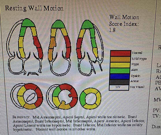
Following bi-atrial maze procedure, the patient converted to sinus rhythm. However, when the cardio pulmonary bypass was reversed and the patient was being closed, he went to ventricular fibrillation followed by defibrillation. Frequent PVCs were observed. Intravenous amiodarone and magnesium were started; His blood pressure started to drop and vasopressors were increased and intra-aortic balloon placed. His hemodynamics improved and was watched in the OR with echo. There was inferior wall hypokinesis. He was maintained on low dose dobutamine, vasopressin and norepinephrine. His global ejection fraction was reasonable; chest was closed, but before he could be transferred out of OR he developed ventricular fibrillation again. His chest was opened and direct cardiac massage and cardioversion done with return of circulation. ECMO (Extracorporeal Membrane Oxygenation) was initiated. He was transferred to the cardiac cath lab and underwent emergency coronary angiography.

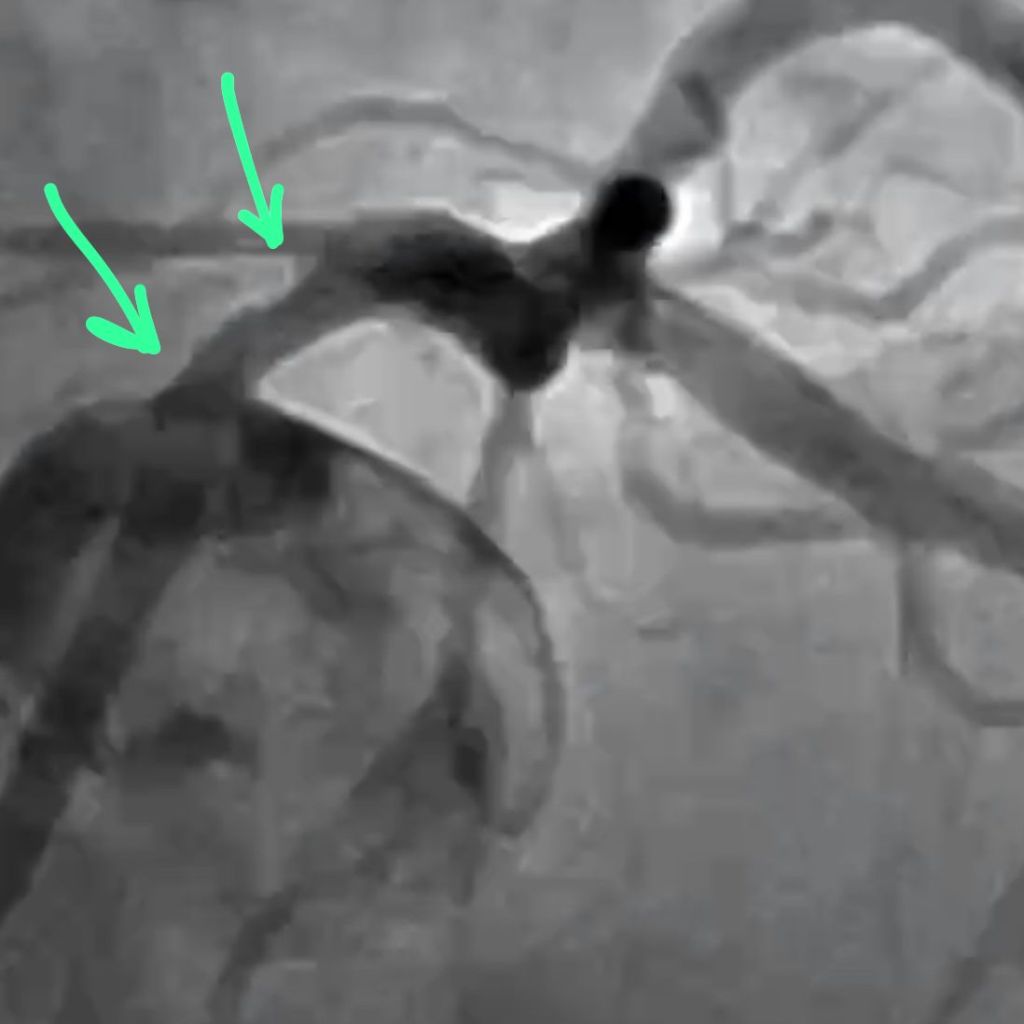
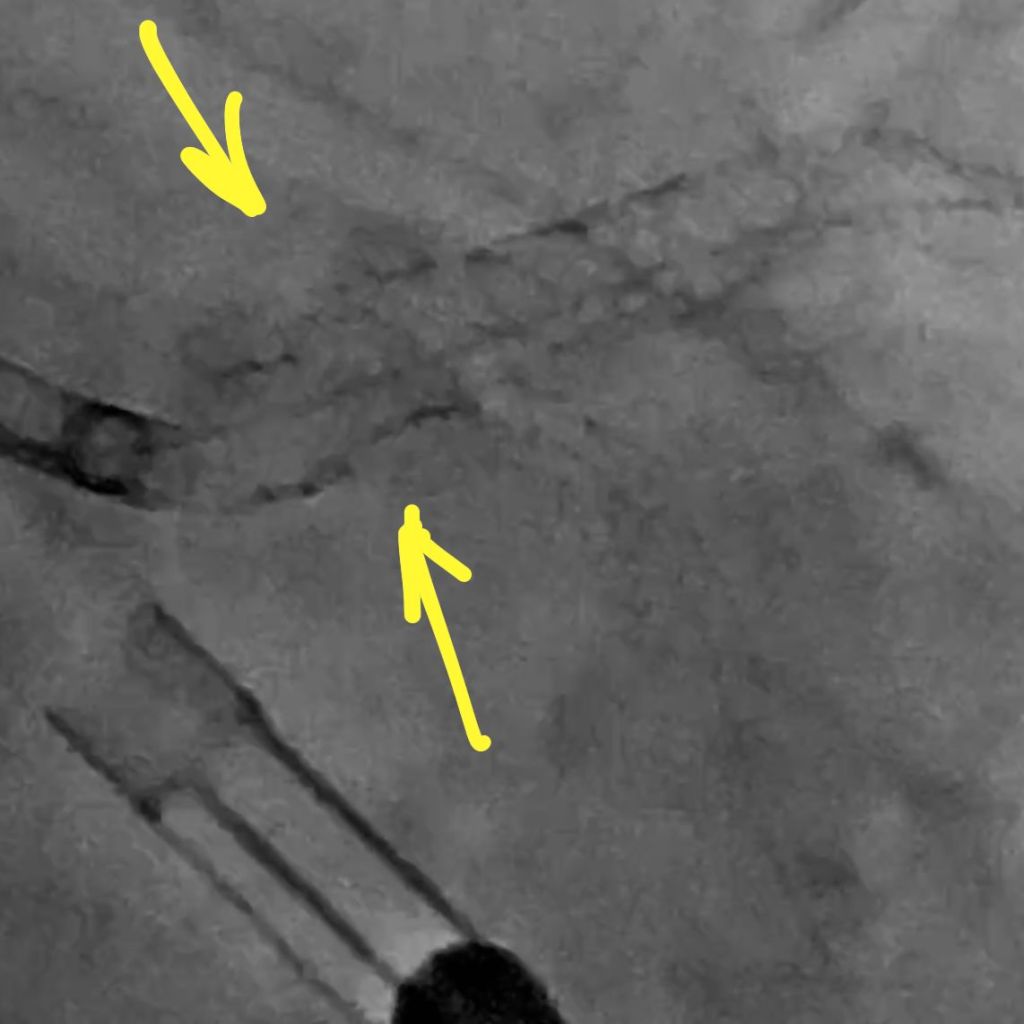
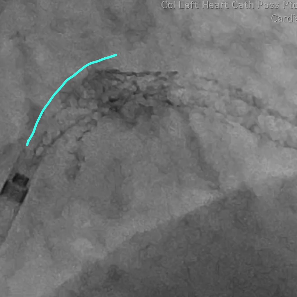
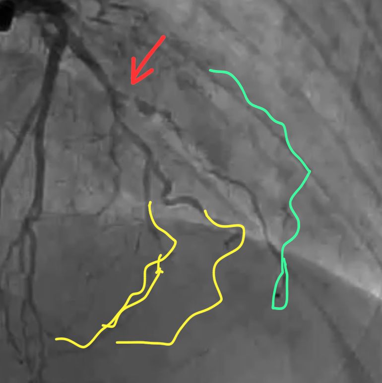
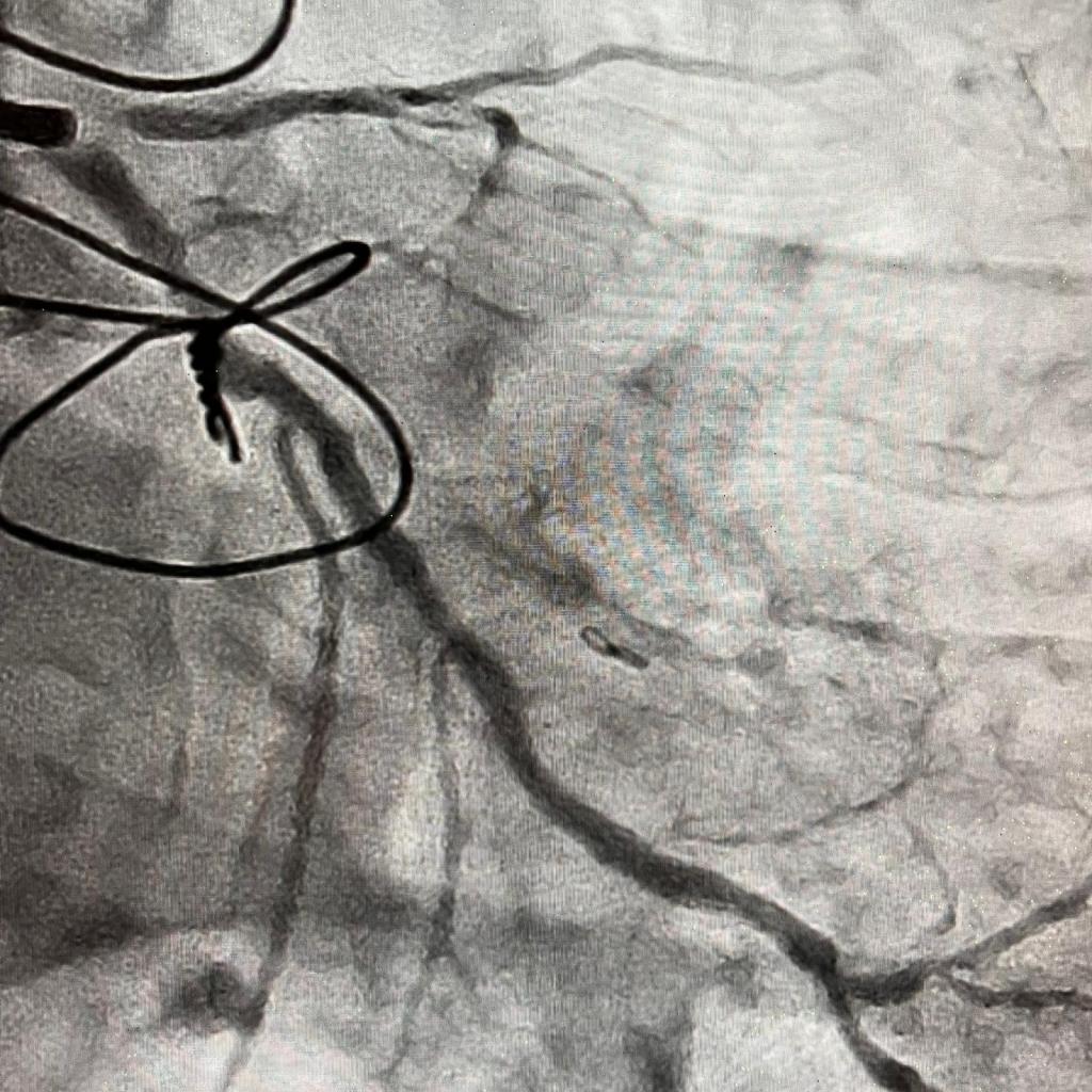
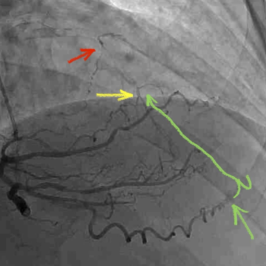
CORONARY INTERVENTION: Coronary angiography showed the dominant left circumflex was occluded in the mid potion.

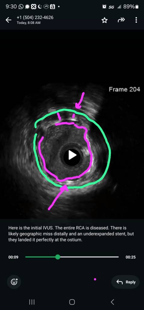
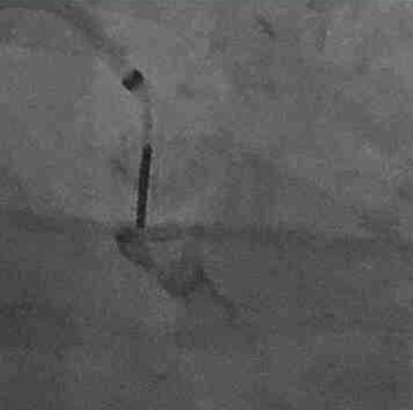
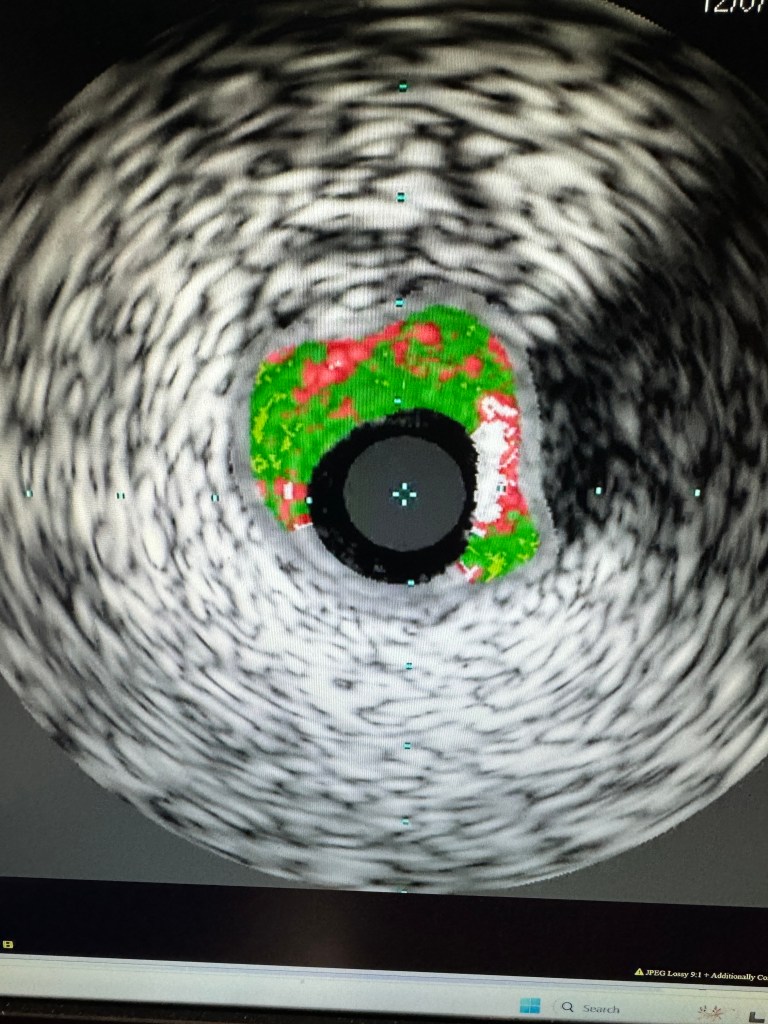
IVUS showed possible edema around the left circumflex and it was felt that the circumflex was occluded due to pressure from the mitral valve ring. Stent was placed with recanalization. Impella was placed in the cath lab and the intra-aortic balloon was removed.

Intavascular Ultrasound (IVUS) shows the edema/hematoma? aeound the left vircumflex artery.


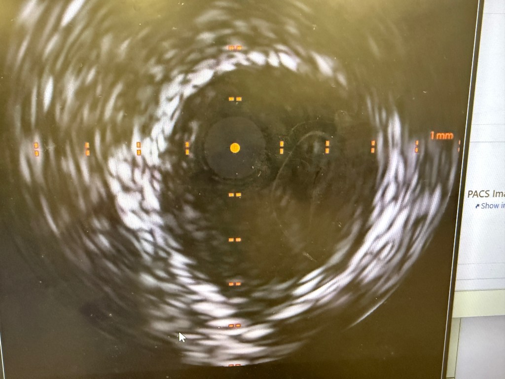
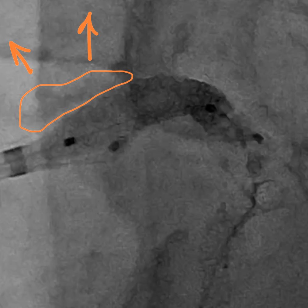
A 3.5 mm x 32 mm Synergy drug eluting stent was placed in the mid circumflex coronary artery with a good result. The intra-aortic balloon was removed.


INTENSIVE CARE UNIT: Patient remained with supported blood pressure, with severe anemia needing multiple blood transfusions. Transesophageal echo showed severely depressed right and left ventricular systolic function.
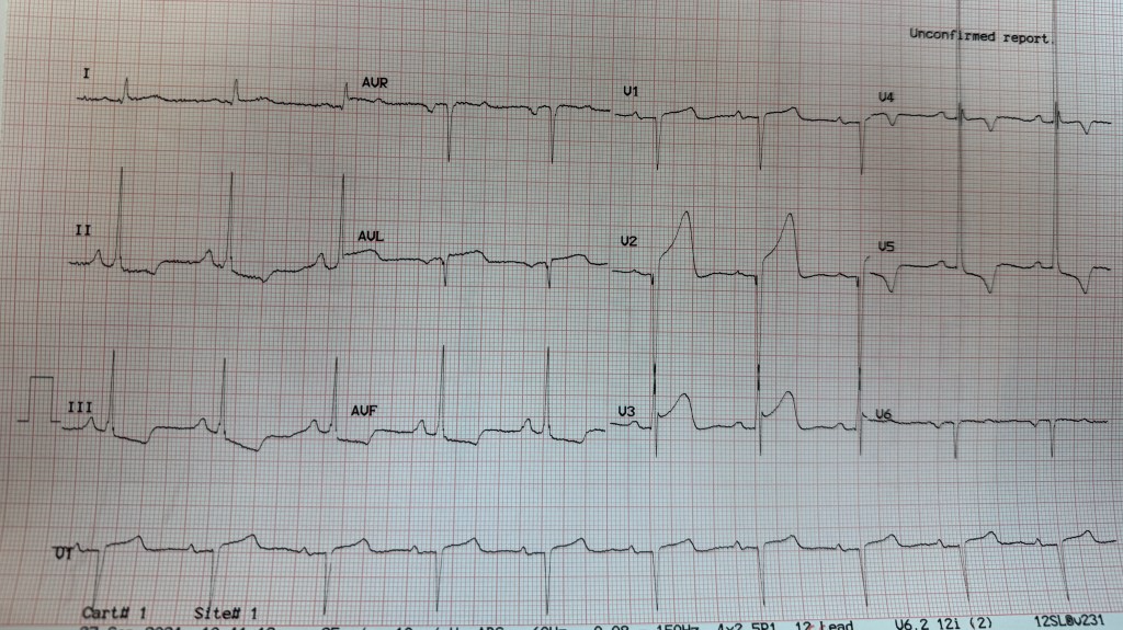
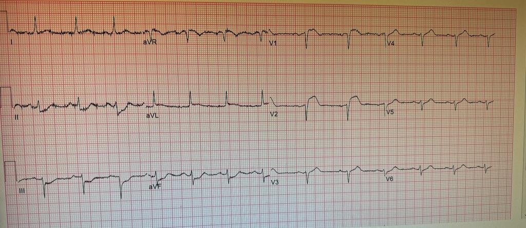
Patient was taken back to O.R. for exploration and washout. On return to ICU noted to have ST elevation in lead VII. Taken back to cath lab and coronary angiography done showing that the circumflex stent was patent.

FOLLOWUP: 10 days after mitral and tricuspid valve repair and coronary intervention, patient is off ECMO, and on Impella support. He is awake but does not follow commands. His global ejection fraction on echocardiogram is mildly depressed (40-45%) with trace of mitral and tricuspid regurgitation.


STEMI on the Forehead !

Amin H. Karim MD
Institute for Academic Medicine
Houston, Texas

Today a 65 year old patient showed up in the office. He had not seen me for last 5 years for whatever reason and now needed a surgical clearance. He worked as a RN at a Houston Hospital and subsequently at the VA Hospital;

I did a routine EKG as follows:

It was unchanged from his previous EKGs from years ago. When he was following with me, he had a cardiac workup including imaging tests showing normal coronaries.

On taking his interval history, I found some interesting facts. While working at the VA he had some atypical chest pain. He went to the ER and had an EKG and was seen by a cardiologist stat. He was rushed to the catheterization lab and had a diagnostic cardiac catheterization which showed normal coronaries. A few months later he had similar chest pain and again went to the ER at a premier hospital in town for evaluation. An EKG was done and STEMI was called immediately. He protested to the cardiologist that he had cardiac catheterization a few months ago and that it was normal! The cardiologist was insistent that he have another one since his EKG showed that he was having a heart attack and could die. His protests were to no avail and he was rushed to the lab and had another diagnostic cardiac study which of course showed normal coronaries. The cardiologist came and apologised to him for not listening to him.

Now it was my turn to beat him on the head and tell him: ” You have STEMI written on your forehead. Wherever, you go you will be treated with emergency response and the STEMI will be summoned and you will be rushed in an elevator with a security guard holding it! Once in the cath lab you will be surrounded by a frantic team counting minutes!

So this is what you are going to do. You will take a copy of your EKG, go to Office Depot and have it reduced and laminated; then carry it with you in your wallet and if you land in the ER and before they wheel you away to the cath lab, show them the EKG and tell them that your EKG is always abnormal and that they should compare to see if there are any changes. Otherwise you will someday end up with a clot in your hand or leg or some other complication!

I think he got the message.

No offence meant and take it in the lighter vein, but I am sure every interventional cardiologist taking emergency calls faces these alarms and the 90 minutes door to balloon time does not allow much margin to hunt for old EKGs!

Amin H. Karim MD
September 27 2024

Foot in the Door: A Complicated RCA!

By Salman Arain MD

Case of the Week – A Foot In The Door May Be All You Need!

Here is a complicated RCA intervention from this week with several discussion points. There is a bonus here – my technique for changing guides over a coronary wire. There are several sequences – but not as many as there could have been!

Baseline angio. 73 year old woman with severe angina – history of bypass with failed RCA graft. Has critical ISR at the RCA ostium. The referring had difficulty stabilizing a JR4 guide and was unable to wire the stenosis.

Here the initial wiring sequence. We used an AL1 to engage the ostium as best as we could. We used a Fielder XT to wire the ISR – it is the perfect wire to do this because of the tapered tip, the polymer jacket and the supportive shaft.

But that AL1 position is not ideal for working. The next is to build up support. We used a TurnPike LP to dilate the channel and switch out for an exchange length wiggle wire. TP LP is ideal because of its double nitinol braid – it can drive itself forward even when support is suboptimal. Once the wire is in, we can dilate with a long balloon. IVUS to follow.

Here is the initial IVUS. The entire RCA is diseased. There is likely geographic miss distally and an underexpanded stent, but they landed it perfectly at the ostium. An AL1 is not the best guide for the remaining sequence which involves engaging and disengaging the RCA ostium. So we decided to switch guides.

Here the entire sequence to switch the guides over a coronary wire. You need a long (preferably supportive) coronary wire, and exchange length 0.035 wire, and patience!!! The 0.035 wire straightens out the AL1 during transfer.

Here is the post IVL result by IVUS. The calcium ring is disrupted is several places!

After using a 4 mm NC balloon, we implanted a 4 x 48 mm SYNERGY. Here is the post stent IVUS. We post dilated with a 4 mm NC balloon.

Here is the final result! You can see the contrast in size between the stented segment and the native, diseased RCA. There is always a risk of ‘over stretching’ the vessel when IVUS measurements are strictly followed, but this is an acceptable ’step down’.

QUESTIONS:
AA Salman bhai, I had 3 questions for my own learning, not questioning. Angiographically its a beautiful result as expected from you.

is the stent truly undersized or just underexpanded (heavy calcium) in some areas by the original operator?

any for shockwave or laser (not great for calcium ) to help expand the calcium. on your post ivus the calcium appears to prevent expansion but angiographically looks awesome. risk (calcium perf ) /reward of expanding this area with a short 4.25 NC given his prior restenosis and ostial location. i would have left it as you did,

why wiggle wire? what other wire if not wiggle wire?

Good questions.

1) Undersized versus under expanded? Difficult to say. As you know, most stents between 2.5 and 3.5 mm are the same size. The difference is in the size of the balloon that they are mounted on. (@Waqas T. Qureshi MD Bhai can confirm or refute this). So, it is difficult to say whether the stent was just undersized from the get-go or not fully expanded. But it is clear that there is geographic miss and under preparation of the vessel.

2) Absolutely. I highly recommend aggressive post dilation after any atherectomy or IVL. You have to ensure the vessel will expand before the deploying another stent.

2b) This also highlights the limitation of angiography alone to gauge vessel size in diffusely diseased arteries.

2c) Post dilation options include noncompliant balloons and (now) OPN.

3) Any supportive wire may have worked. I like to use the wiggle when dealing with torturous RCAs or LADs. The wiggle portion often locks the wire in position, adding to the support provided by the wire. Of course, any other support wire may have also worked.

The Right Ventricular Branch: An Exception to the Rule!

By Dr. Salman Arain
Lately we have had several complex (and complicated) RCA interventions. In such cases we usually don’t worry about the RV branch – if it arises from a diseased segment we protect it, if it away we let it be. Also, the transient loss of an RVB is mostly (but not always) well tolerated. Here is a case that is the ‘exception to the rule’.
Introduction: 75 year old man with known CAD. History of RCA PCI (two?) years ago complicated by perforation and placement of a PTFE covered stent. The patient returns with progressive angina.

Interestingly there is severe ISR within the covered stent – which may explain the rather late presentation. Typical ISR presents within 6 to 12 months.

Our plan was to perform PTCA, hopefully provisionally but you can see the difficulty we had advancing even short balloons. We resolved the support issue with a buddy wire and a guide extender.

There was recoil and we decided to stent with an Orsiro (sirolimus based) stent but we had difficulty in advancing it. The RAO reveals why – a ledge of calcium in the mid RCA! We took care of it with lithotripsy (Shockwave).
Shortly after 10 placement, the patient started to complain of chest pain. He also had diffuse ST depression. We repeated the angiogram, but the flow looked good.

We admitted him to the CCU where he had a modest increase in his cardiac enzymes. The high sensitivity troponin went as high as 18,000(!). The levels started to come down on day 2.

The patient continued to complain of exertional jaw pain on day 3. We maximized his antianginals as much as a blood pressure allow, but he continued to be symptomatic. The echo showed normal LV function.

We took the patient back to the lab and found that his RCA was still patent. There is TIMI-2 flow possibly due to the recent PCI and large vessel diameter. However, we can see that there is a small RV branch that had a) disappeared during the first procedure and b) is trying to come back. We decided to open “”rescue it”.

However, this is where things became ‘interesting’. We had considerable difficulty engaging the jailed RVB ostium with multiple wires (both with and without a microcatheter). You can see the challenges here, which include lack of support and a predisposition for wire prolapse.
Some additional thoughts:
1) We took a picture of the left system and it was unchnaged from 3 days ago

2) There were several challenges in wiring: the ostium was jailed, embedded in disrupted calcium, retroflexed or at least perpendicular to the MB, and a relatively wide RCA lumen.
I tried multiple polymer jacketed (slippery) wires to no effect. I even tried a Fighter (0.008 tip) from Boston because it has a non-jacketed tip. For a different grip. But it did not work. With the blocking balloon technique, the wires just kept curling up in the RCA. Also, having a new stent struts did not help – too much exposed metal.
Case Resolution: The Micro JR4!
We decided to try a SuperCross 90 (angle tipped catheter). I gave it a small secondary bend to create a micro JR4, and to our pleasant surprise, it was able to engage the RVB ostium!

Here you can see the micro JR4 in action! We used an anchor balloon to drive the TP LP across the ostium, thus dilating it.

This then allowed us to insert a 1.5 and then a 2 mm ballon. The final angio shows brisk antegrade flow in the recovered RVB.
The patient’s angina resolved completely after the procedure. He felt great and wanted to go home the same day, but we kept him overnight just in case.

An interesting observation: The collateral from the LAD is what kept the distribution of the RVB alive. That is why he continued to have angina, even after the enzymes plateaued. I think it took a day for this collateral to plump up, which is why he suffered immediately after the branch went down.

THE END
What a nice case 👏👏 , masterfully done Salman bhai.
questions
1- would atherectomy have had a lower chance of shutting down side branch compared to lithotripsy
2- did you KISS or POT the RCA after this final ballooning or not needed ?

1) Possibly. Had we used atherectomy, we may have had less disruption at the RV ostium, and we would have cleaved the calcific plaque.

2) I don’t think we did. (Someone else asked and I said yes, but when I checked I couldn’t find the clip).

The goal of the SB balloon was to open the ostium and stretch the struts. Hopefully, the perpendicular takeoff minimized carina shift into the RCA proper.
Great questions Waleed Kayani Bhai. Sometimes we do things “in the heat of the moment” but looking at the case again (alone and with colleagues (like the Houston Cardios!) opens up other possibilities. 😀


Evolving Nature of a Complex Bifurcation PCI

By Dr. Salman Arain

85 year old man admitted with CP in setting of HTN. Had has symptoms for a few months. ECG showed ST depression and the EF was 45%. The referring sent him for CABG but the (chief) surgeon was hesitant because the patient is a Jehovah’s witness (can not receive blood), low platelets (85k), and high Cr (1.9)

.

What is your ABC analysis? As a reminder:
A – angiographic analysis,
B – bifurcation strategy,
C – calcium modulation,
D – device (MCS) selection if any, and
E – execution strategy?

Ok, so the resolution of our case…

These were my thoughts going into the case…

A. Functional last conduit because the RCA is critical. The entire body of the LM is involved. Heavy calcium is an understatement.

B. Medina 1,1,1 – both arteries need to be addressed. Our plan was to perform DK crush.

C. Rota and/or IVL. ‘Relative’ disadvantages to both – Rota will require me to remove the wire from one of the branches. IVL will prolong LM occlusion (= ischemic) time.

D. Definitely Impella supported.

E. Plan – perform RHC for reserve, insert Impella, wire both LAD and LCX, PTCA LM into LAD, IVUS then decide on rota strategy, finish with DK crush. Stage RCA.

We started by doing a RHC. The PA pressures were not too bad (48) and the PA sat was reassuring 58%. We opted for dual access.

We resolved that issue with a TurnPike LP microcatheter. You can drive the MC by torquing it. We knew we were going to fix the LAD with rota so I did a 1.5 mm angioplasty to allow the LAD to breathe while we did IVUS.

With the LAD and LCX fixed, the LM is more manageable now. We opted to finish the T-stent and use IVUS to see if further optimization was needed.Our bifurcation plan rapidly evolved during the case. It was DK crush before the case, mini crush after I put the proximal LCX stent in, and ended up being a T-stent because of how well the stent had landed at the ostium.

You can see the patient is becoming restless. I think that it was likely from leg ischemia related to the Impella sheath.

Also, the guide had softened and came out. Note the stent deformation on account of the calcium module at the ostium. There is a recoil after high-pressure NC ballooning.

Here is the CFA after Impella removal. We did not use single access because I wanted to use a 7 French guide without any of the drama. A 7 French sheath is often quite snug in the Impella insertion sheath. The double access also allowed us to ensure that the bleeding at the Impella insertion site had completely stopped before the patient left the Cath Lab. This was on on account of his inability to receive blood being a Jehovah’s witness.

The patient did very well. He was up and about the next day. He did have a mild bump in his creatinine, but we kept him for a few days.

I plan to perform an PET in four weeks and then decide if he needs the RCA fixed. Again, he is an 85 year old who has been doing quite well until now.

The End. 🙏🏼

I am going to share a series of still frames that shows the changing morphology of the LM lumen. This is baseline.

Here is the balloon inflation. Note the upward displacement of the nodules.

This is after our aggressive POT. Note the smoother upper curvature.

The Gordian Knot

Using Contrast To Cut The Gordian Knot! A Complicated CTO Intervention

By Dr. Salman Arain

This is the complete angio. The patient is 57 years old with CCS 3 angina. He had bypass surgery 13 years ago. His EF is 35 to 40%.

Viability Study

So, the question for our Cardio experts:

1) What do you recommend? PCI, re-do CABG, EECP or OMT alone?
2) If you choose PCI, which vessel would you go after first?
3) And what CTO strategy would you use? Retrograde PCI may or may not be the best answer here.
4) What do you know about the coronary sinus reducer, and would you consider that?

Tricky. The problem with the LCX is that the caps are all ambiguous and there are several bifurcations.

The LAD CTO is long but at least there is a well defined cap and a relatively straight course. Unfortunately the LAD septals supply the PDA which itself is occluded proximally and does not connect to the PLBs.

The RCA is the trickiest of all three. One thing to keep in mind is that the patient is post CABG – so any perforations would be more difficult to treat.

Ok, here is how it all played out. First a few thoughts about choosing the vessel to intervene upon.

The LAD has the most favorable anatomy but needs to be fixed least urgently. The proximal 1/3 is patent and the mid segment has a graft.

The LCX has an ambiguous cap and several branches that need to be rescued. It is important not only because of its own distribution, but because it is the best retrograde conduit to the RCA via the PLB.

RCA is the trickiest because it requires a combined antegrade and retrograde approach. Also, the PDA is occluded and is cut off from the large PLB system. So an occluded bifurcation somewhere!

Thus, we decided to go after the LCX – it was the one that would give us the greatest advantage in addressing the RCA.

Here are some dual injection angios to better show you the connections.

There are several ways to tackle a CTO – some antegrade and some retrograde. We have focused on and specialized in contrast modulation of plaque. This requires puncturing the proximal cap and then using micro injections of contrast. The technique is also called a modified Carlino injection after Mauro Carlino who described

Here is the setup for the LCX intervention by means of CAP (aka Carlino). Notice the ambiguous cap.

This is not the typical cloud. It is tubular which means we must be extra plaque

So we used a Gaia 2 to redirect our MC and repeat the contrast injection. Now we see the vessel architecture – as well as re-entry!

This is just the injection clip. The typical result is seen towards the LCX. There contrast mediated STAR into the OM2!

So we wired the LCX first, then treated the bifurcation, and used a dual lumen catheter to wire the OM2.

We used IVUS to confirm that we were not going to jail off the side branch with our stent. We used JSBT!

And here is the final angio. You can see that we are nicely set up for a retrograde intervention on the RCA in a few weeks.

And now you have the complete name for the case: Using Contrast To Cut The Gordian Knot! A Complicated CTO Intervention

Afterword. I think this case highlights how far coronary PCI has come – as a specialty. In my fellowship, this talk of dissecting and injecting contrast and going epicardial would have been blasphemous. And now it is routine in selected centers of course.

The upshot is that CABG is no longer the end of the road. The end of the road is the end of the road – and that is not in sight for coronary PCI!!!

Dr. Zaka Khan wrote: Regarding Coronary sinus reducer – This was original developed by Neovasc . A Canadian startup. There main two products for Cosira and Tiara. Cosira was designed for controlled occlusion of CS to improve symptoms of refractory Angina . It does improve at least 1 CCS class. Would work only in cases of refractory angina and evidence of reversible ischemia. It increases coronary microvasculature flow.
Tiara was supposed to be Transseptal Mitral valve replacement system without need for apical approach.
Company got into litigation with Edward’s and then few guys at UPEN. Material used for Tiara was manufactured by Boston Sci and hence they jumped into the mix. Cosira system was bought into by Shockwave medical.
Tiara is still somewhere and the fight goes on.
Many investors lost money because of legal battles. Would have been a big hit like TAVR .

Questions by Dr. Afaq Motiwala answered by Dr. Arain:

) Does the micro catheter injection into the cap create more extensive dissection (if you are extra plaque) and make it harder to get into true lumen? Yes and no. That is where we have honed our technique. It’s all a matter of understanding the contrast cloud.

2) How did the ivus help you with side branch? It helped confirm that I was in true lumen and that the side branch was not jailed behind plaque.

3) Where did you place your balloon for jsbt? Balloon from OM2 to top branch and stent in the main body of the OM2

4)The small branch you preserved with a wire, does it have some contained hematoma? Any need to tamponade it or it’s inconsequential? It does, but the flow was good without a dissection in multiple views. So OK to follow clinically. These are very small branches.

.

It was important to preserve the OM2 trifurcation. You can see the size of it in the cranial view. The top branch fills OM1 and the bottom two supply the PLB. I plan to fix both eventually.

Dr. Amin H. Karim wrote: The cath diagram took me back to pre-EMR days when we routinely made these after watching the cine on Trajano. They were very reflective of the coronary anatomy as well as recording one’s personal notes. 30 years later when we look at these in the patient’s chart it gives a good idea of the pre-intervention anatomy. Computer diagrams are no match and I feel sorry for your younger colleagues when they pull their patient records decades later, they will be looking at a computer cartoon that is a far cry from the real anatomy. Both Michael DeBakey and Denton Cooley gave a lot of importance to the cath diagram on patients referred for surgery and both would hang them on the old x-ray box in the OR as a reference! Dr. DeBakey would meticulously put his pencil notes on the diagram and file them on each patient. He would also draw the grafts on another copy and send them to the referral cardiologist. He would pull the file out when patient came in years later for redo

How to Plan A Left Main PCI – The ABCs

By Dr. Salman Arain MD

Here is a way to think (and talk) about left main PCI.

A – Anatomy (i.e. functional anatomy) which includes angiography, IVUS, and physiology.
B – Bifurcation classification If applicable of course.
C – Calcium management. All inclusive term that includes all available devices for lesion preparation: rota, CSI, IVL, cutting balloons, laser, and of course NC balloons.
D – Devices. This includes the need for and choice of mechanical support.
E – Execution. The actual PCI strategy (or strategies).

86 year old man with patent LIMA and SVG to RCA. Now with exertional angina and lateral wall ischemia (large).

Here is the LIMA shot from then – the proximal segment of the LAD is not supplied by the graft. So worth saving and/or protecting.

Here are my ABCDEs – as promised.

A: Functionally and anatomically the calcium involves all three branches. FFR or iFR not needed.

B: This is a Medina 1,1,1. The proximal LAD supplies a significant area and is worth saving.

C: There is nodular calcium and some type of atherectomy will help place stents. My first choice was rotational atherectomy given that it is a front cutter.

D: This is a protected LM and the EF is low normal. So no need for an MCS.

E: The LM is very short or non-existent. This anatomy is better suited for Culotte though any technique would work.

S: Not applicable.

We ended up wiring the LM into the LCX followed by atherectomy with a 1.5 burr. We were unable to wire the proximal LAD from the LM because the ostium was obstructed by the calcium nodule.

And here is the final result.

You can see that the LCX is technically a first OM that supplies a major portion of the lateral wall with its 5(!) branches.

A Tough Chronic Total Occlusion (CTO)

By Dr. Salman Arain
McGovern Medical School, Houston, Texas.
February 10 2024

There are several ways to approach a CTO. The entire crossing process, whether antegrade or retrograde, starts with crossing the cap. Different techniques have been described. In my practice, I like to use contrast to modify and modulate the cap/plaque…

History. 68 year old man with LAD PCI 4 years ago. Presented with angina and found to have a new RCA lesion (which was stented) and this LAD CTO. The angina has resolved but the exertional dyspnea persists. The PET was very abnormal in the AW.

There are 3 sets of right to left collaterals. Not all are ‘usable’. The LAD fills in ‘stages’ which suggests disease in the native LAD beyond the CTO.

Plaque penetration was more challenging than expected. That is a Hornet 14 wire (14 gm penetration force at the tip!). As per our protocol, we stopped once the wire was a few mm distal to the hard cap. We then maneuvered our microcatheter (MC) in place.

Contrast modulation. 👆🏼Here is the intra-CTO contrast injection! Note the free passage of an tapered tip hydrophilic wire (Fielder XT).


Here is a distal tip injection. This is different than injecting into the occlusion. Transducing pressure allows is to confirm we are across before injecting. A softer tipped hydrophilic wire (Gladius Mongo) helped us track into the true LAD.


I like to perform IVUS before stenting to understand the nature of the CTO, the characteristics of the vessel wall, and the size of the lumen. This helps us choose the appropriate vessel preparation and stent therapy. Here our crossing is all intraluminal.

Here is the final run. We did multiple IVUS runs to size the LAD and optimize both the LAD and the D2. Note that all septals have been preserved! All in all a successful CTO which we completed in under an hour and a half.


The Steel Belted LAD

by Amin H. Karim MD

A 75 year old Caucasian female with HTN and Type II Diabetes Mellitus was admitted with few hours of chest pain. EG showed ST elevation in V1V2V3. STEMI team was called. She had no previous history of CAD. Had a negative EKG stress test just 6 months ago.


It took thrombus aspiration with Export catheter, IVUS, and placement of 3 stents totaling 80 mm to achieve this result

EKG stress test is not a reliable predictor of adverse coronary events. it can miss even severe coronary occlusive disease, as demonstrated above; Coronary Calcium score may have increased the sensitivity. The right coronary artery will be tackled two days after the myocardial infarction.

2 days after the LAD intervention, we tackled the RCA again requiring multiple stents to treat the multiple focal lesions; a total of 96 mm of stents were placed in the RCA.

Beware of the Bridge

By Amin H. Karim MD
Joseph Aaon MD (Interventional fellow)
We present a case of acute myocardial infarction with total occlusion of the left anterior descending coronary artery. A 85 year old male presented with several hours of atypical chest pain. EKG at a free standing ER showed ST elevation in the antero-septal leads. Catheterization was done in the 90 minute door to balloon time and showed the following: There was a vessel alongside of the left anterior descending coronary that looked like a bridging collateral. Our question was: is this a chronic occlusion with a collateral or was it an acute occlusion causing the chest pain and EKG changes. Orthogonal views were obtained to make sure we do not end up wiring the bridging collateral, which may give the impression of wiring the true lumen. hence it may not hurt to obtain one or more orthogonal views to confirm the position of the wire. The micro catheter also helps if there is confusion. Injection of the contrast into the vessel can differentiate a bridging collateral from a branch of the vessel. 

The lesion was crossed using a micro catheter and a whisper wire. 3.5 x 38 mm DES was placed. Final result was satisfactory. 

IVUS pre dilatation showed a fair amount of calcium. 

Echocardiogram obtained the next day showed anterior wall and apical akinesis. There was a layered thrombus in the apex of the left ventricle. Patient was anticoagulated with warfarin, in addition to clopidogrel and aspirin. Plan was to discontinue aspirin in 2 weeks to reduce the risk of bleeding.