Challenge #1. Here is the baseline angio. The challenge here is wiring the mid LAD. The wire tip shape needed to cross it is different than the one needed to reach it. Our solution – a dual lumen catheter (Sasuke) in the mid LAD which allows us to secure the D2.
Challenge #2. We wired the D1 to secure it during provisional LAD stenting. There is plaque shift +/- thrombus which shuts down the D1 by the time we have treated the mid LAD. Luckily we still had the wire in place – so in goes the Sasuke again. We were able to guarantee that the wires did not end up behind the stent struts.
Challenge #3. We did a proximal POT, pulled the jailed wire, and passed it down the LAD. I did not want to keep working on the Fielder. Note the brief detour into D2. Alas, we ended up with a dissection! After this point the patient started vomiting and the ST were really deep. I felt that the 6 Fr guide was workkng against us. As you can see much of the LAD has shut down. Our solution – intubate, insert Impella, upsize to 7 Fr.
Challenge #4. Here is the groin angio through the 14 Fr Impella sheath. The leg will likely become ischemic during the case- so we put in a retrograde SFA sheath for future ‘external bypass’.
Challenge #5. Our first angio after Impella placement shows clot in the entire LAD!!! 😳😩 I called for Penumbra, but decided to make a pass with an Export while the cath lab was setting up. Surprisingly we were able to ‘uncork’ the LAD.
Challenge #6. The next step is to treat the D2 and optimize the mid LAD stent. There is stent recoil. IVUS showed a fibrotic lesion – we treated this with a cutting balloon and completed the LAD PCI. Or did we…
Challenge #7. The completion angio shows thrombus in the proximal LAD – address it with medications or aspirate? We tried PTCA but it embolized. Well, by this time the Penumbra was set up. We were able to complete the procedure (again!). This is the final coronary angio.
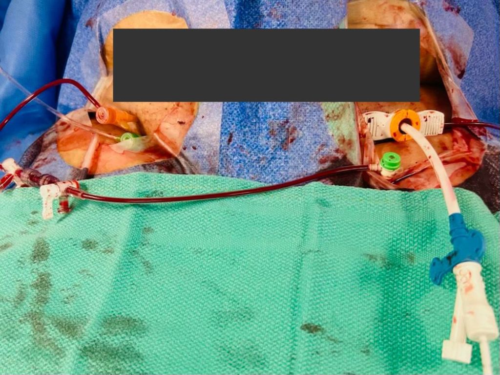
Challenge #8 (No more, I promise): How to keep the leg perfused while the Impella was in place? Here is a clip of the initial angio through the Impella sheath. The third clip is through the SFA sheath (using a micropuncture dilator) after the external bypass has been set up (right CFA -> left SFA). I do this to confirm pulsatility in the occluded leg.
This is what the set up looks like. There are several advantages to doing it this way. The retrograde sheath is easier to insert, doesn’t compromise antegrade flow to the leg, allows you to do an angio to confirm Impella sheath closure (if done), perform internal balloon tamponade on the proximal arteriotomy (if needed) and importantly – makes sure there is no dead zone in the CFA. As a bonus, you can close this with an AngioSeal later if needed.
The End. Question: How would you take out the antegrade sheath? Manual and balloon tamponade the CFA from contralateral? Answer: They are both retrograde. The angio shows it. It looks antegrade because of the body habitus. The typical sequence is: 1) Remove Impella 2) Exchange Impella sheath for new sheath without flushing 3) You may do an angio from the lower (SFA) sheath at this stage. This is optional. 4) Treat the proximal arteriotomy with Manta or double Perclose. 5) Confirm closure with injection through the lower sheath. 5a) Consider balloon tamponade from the lower sheath if needed. 6) Close the lower arteriotomy if the SFA is ‘clean’ – you must use a device. It is too deep for manual hemostasis. 7) Confirm close from the contralateral side.
1) These are recommendations for Impella inserted in an emergency – and expected to stay in for some time. Usually there is no time for Preclose if the patient is crashing. 2) Also, I don’t Preclose unless I know the Impella is coming out within 6-12 hours. 2b) We have left the sutures in longer but management becomes an issue if the CCU stay is prolonged. 3) You don’t have to put a new Impella sheath in. Any 14 Fr sheath will do. 4) The idea is that there may be thrombus in the old sheath – in the space between the sheath and the Impella. 4b) Of course, you can hook it up to pressure to keep it open – but we have had thrombus form despite that too. 5) When the Impella comes out, you can use your favorite method to obtain hemostasis. Double Perclose is just one. 6) Stick the SFA in the cleanest part – avoid being too close to the Impella sheath. You don’t want the lower sheath tip ‘tucked under’ the Impella sheath.
We have left Impellas in upto 5 days with good leg perfusion using this method. The best part is that you can take it out yourself in the cath lab. No need for the OR. Question: SA; trying to understand.. The need for# 6? Why is the SFA stick needed? This is required when the Impella sheath is occlusive in the iliacs or the CFA. In such cases the leg with the Impella becomes ischemic.
One way to prevent this is to gain antegrade access into the CFA or SFA and create an ‘external bypass’. Usually this is done with US guidance and is a little tricky.
Our method makes it wasy because you can stick the SFA without changing the side you are standing on. Also, if you use a roadmap (like we did) you can do it without US. Because there is usually some flow via the profunda: CIA -> IIA -> PFA -> CFA/SFA.
Question: Also, for the SFA are you using an Arrow sheath ? Answer: Yes, always an Arrow sheath. Given the depth of the vessel and the angles involved, you need a braided sheath. Non-braided sheaths will typically soften and kink.
By Syed Aman Ali Medical Student, Jinnah Sindh Medical University, Karachi. Pakistan Amin H. Karim MD, Houston, Texas
A condition that, once diagnosed in a young person, can be treated conservatively with diet and rest, can end up being treated expensively in a milieu where defensive medicine and financial incentive join together. The case below is an example.
Case Presentation
A 20-year-old male student presented to the clinic with a 1-week history of abdominal pain and jaundice. He described the abdominal pain as a heaviness localized to the upper right quadrant, rating it 8 out of 10 on the pain scale. The patient also experienced mild fever, vomiting, and a headache. Patient later noticed jaundice, evidenced by icteric eyes and yellowing of the skin. He also reported dark urine and pale stools. He had recently traveled from Pakistan to the United States and went on a cruise approximately 2 weeks prior to the onset of symptoms.
On physical examination, the patient appeared generally well but jaundiced. Examination of the abdomen revealed tenderness in the upper right quadrant but no guarding and an enlarged liver. Patient was advised bed rest and symptomatic treatment till he felt better and was sent home.
Lab results were returned they showed that Hep A antibody was found to be reactive while Hep B and Hep C were non- reactive. Patient had a high total bilirubin of 12.8 mg/dl and alkaline phosphatase was raised to 182 U/L. AST was raised to 4317 U/L and ALT was raised to 5340 U/L. Patient had an increased hemoglobin 17.8g/dl and hematocrit 55.9%. The plan from our end continued to be conservative and symptomatic. At the insistence of parents, patient saw a gastroenterologist for a second opinion and was immediately sent to the emergency room where he was admitted to the hospital for three days. A second gastroenterologist was called upon to see the patient. All of the lab reports were repeated and more tests were done including Epstein Barr virus test, cytomegalovirus test, thyroid panel. Patient had an ultrasound of the abdomen showing normal results an abdominal CAT scan showing normal result to be followed by an MRI of the abdomen which was also normal. The rationale for doing all three tests with a low pre-test probability of finding anything of significance was not known, Liver function tests were repeated on a daily basis. Liver biopsy was contemplated but not done. Family was reassured by providers that all is being done to make sure the condition does not become worse and that no “other conditions” are being missed!
In summary, the overall cost for the patient’s treatment, considering all expenses, ranged from $20,000 to $24,000. This cost reflects the comprehensive management of a benign condition easily treated with supportive care.
Discussion
Hepatitis A is an acute viral infection caused by the Hepatitis A virus (HAV), transmitted primarily through contaminated food or water. This positive-sense, single-stranded RNA virus, belonging to the Picornaviridae family, primarily affects the liver. It is a significant global health issue, especially in areas with poor sanitation. Typical symptoms include jaundice, fever, abdominal pain, and fatigue. While often self-limiting, Hepatitis A can lead to serious complications in some cases.
Complications and Variants
Cholestatic Hepatitis A: Characterized by prolonged jaundice and impaired bile excretion, leading to darker urine and pale stools. Recovery is often longer and more intense.
Prolonged Hepatitis A: Symptoms such as fatigue and jaundice extend beyond the usual acute phase, requiring extended care.
Relapsing Hepatitis A: Involves periods of improvement followed by recurring symptoms like jaundice and abdominal pain, complicating the clinical course.
The chances of the above complications not withstanding, the condition in young people is benign and self limiting with no sequalae, in fact long term resistance to repeat infection
An important factor is to enhance the protocols for diagnosis and therapy. Better resource management and lower total costs can be achieved by establishing standardized care standards that prioritize evidence-based, economical therapies and simplify diagnostic tests to prevent redundancy. Improving the infrastructure for healthcare is also essential. By investing in sanitation and hygiene improvements in high-risk areas, as well as expanding access to early treatment and preventive care through community health centers and mobile clinics, outbreaks can be avoided and the financial strain on the healthcare system can be minimized.
Lastly, encouraging innovation and research can lead to advancements in prevention and treatment. Encouraging research into new, cost-effective management strategies and adopting best practices based on research findings will contribute to better healthcare outcomes. By implementing these strategies into practice, we can improve patient care while lowering costs in a more effective and efficient healthcare system.
Conclusion
In conclusion, this case study reveals a significant and somewhat ironic truth: treating Hepatitis A, a condition that often resolves on its own with minimal intervention, can still come with a hefty price tag of $20,000 to $24,000 in a milieu of defensive cum financially incentivized medical care. To address this, we should focus on preventive measures like vaccination and improved sanitation, which can help reduce both the incidence of Hepatitis A and the associated treatment costs. Additionally, refining diagnostic and treatment practices, investing in better healthcare infrastructure, and encouraging innovation in care strategies can lead to more efficient use of resources and reduced costs. By making these changes, we can enhance patient care and alleviate the financial strain on the healthcare system.
By Dr. Salman Arain Lately we have had several complex (and complicated) RCA interventions. In such cases we usually don’t worry about the RV branch – if it arises from a diseased segment we protect it, if it away we let it be. Also, the transient loss of an RVB is mostly (but not always) well tolerated. Here is a case that is the ‘exception to the rule’. Introduction: 75 year old man with known CAD. History of RCA PCI (two?) years ago complicated by perforation and placement of a PTFE covered stent. The patient returns with progressive angina.
Interestingly there is severe ISR within the covered stent – which may explain the rather late presentation. Typical ISR presents within 6 to 12 months.
Our plan was to perform PTCA, hopefully provisionally but you can see the difficulty we had advancing even short balloons. We resolved the support issue with a buddy wire and a guide extender.
There was recoil and we decided to stent with an Orsiro (sirolimus based) stent but we had difficulty in advancing it. The RAO reveals why – a ledge of calcium in the mid RCA! We took care of it with lithotripsy (Shockwave). Shortly after 10 placement, the patient started to complain of chest pain. He also had diffuse ST depression. We repeated the angiogram, but the flow looked good.
We admitted him to the CCU where he had a modest increase in his cardiac enzymes. The high sensitivity troponin went as high as 18,000(!). The levels started to come down on day 2.
The patient continued to complain of exertional jaw pain on day 3. We maximized his antianginals as much as a blood pressure allow, but he continued to be symptomatic. The echo showed normal LV function.
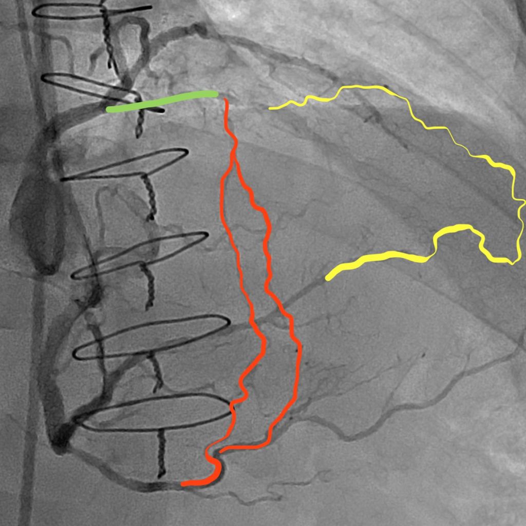
We took the patient back to the lab and found that his RCA was still patent. There is TIMI-2 flow possibly due to the recent PCI and large vessel diameter. However, we can see that there is a small RV branch that had a) disappeared during the first procedure and b) is trying to come back. We decided to open “”rescue it”.
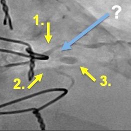
However, this is where things became ‘interesting’. We had considerable difficulty engaging the jailed RVB ostium with multiple wires (both with and without a microcatheter). You can see the challenges here, which include lack of support and a predisposition for wire prolapse. Some additional thoughts: 1) We took a picture of the left system and it was unchnaged from 3 days ago
2) There were several challenges in wiring: the ostium was jailed, embedded in disrupted calcium, retroflexed or at least perpendicular to the MB, and a relatively wide RCA lumen. I tried multiple polymer jacketed (slippery) wires to no effect. I even tried a Fighter (0.008 tip) from Boston because it has a non-jacketed tip. For a different grip. But it did not work. With the blocking balloon technique, the wires just kept curling up in the RCA. Also, having a new stent struts did not help – too much exposed metal. Case Resolution: The Micro JR4! We decided to try a SuperCross 90 (angle tipped catheter). I gave it a small secondary bend to create a micro JR4, and to our pleasant surprise, it was able to engage the RVB ostium!
Here you can see the micro JR4 in action! We used an anchor balloon to drive the TP LP across the ostium, thus dilating it.
This then allowed us to insert a 1.5 and then a 2 mm ballon. The final angio shows brisk antegrade flow in the recovered RVB. The patient’s angina resolved completely after the procedure. He felt great and wanted to go home the same day, but we kept him overnight just in case.
An interesting observation: The collateral from the LAD is what kept the distribution of the RVB alive. That is why he continued to have angina, even after the enzymes plateaued. I think it took a day for this collateral to plump up, which is why he suffered immediately after the branch went down.
THE END What a nice case 👏👏 , masterfully done Salman bhai. questions 1- would atherectomy have had a lower chance of shutting down side branch compared to lithotripsy 2- did you KISS or POT the RCA after this final ballooning or not needed ?
1) Possibly. Had we used atherectomy, we may have had less disruption at the RV ostium, and we would have cleaved the calcific plaque.
2) I don’t think we did. (Someone else asked and I said yes, but when I checked I couldn’t find the clip).
The goal of the SB balloon was to open the ostium and stretch the struts. Hopefully, the perpendicular takeoff minimized carina shift into the RCA proper. Great questions Waleed Kayani Bhai. Sometimes we do things “in the heat of the moment” but looking at the case again (alone and with colleagues (like the Houston Cardios!) opens up other possibilities. 😀
Here is a case I recently presented at CVI. I broke it up into different videos for teaching. A PDF of the complete presentation is included at the end.
62 year old man with HTN, DM2, and CKD 3. Presents with progressive angina for three months. Now CCS class 3. The referring MD sent him for a CTA – no stress test available. How would you approach this?
There are several notable features:
Anonymous left main from the non-coronary cusp.
Proximal LAD occlusion with a diseased mid segment.
Bifurcation disease involving the high ramus/OM1.
Patent LIMA, which supplies the distal LAD. 4b. Patent intracostal branch from the LIMA, which may be causing a steel phenomenon.
Moderate disease involving the takeoff of the high PDA.
Occluded SVG to the PDA (not shown).
As such, it is difficult to determine the exact location of the ischemia. There is also a second diagonal branch, which is diffusely diseased and supplied by epicardial collaterals.
I asked the referring MD to get a stress test. The patient had ischemia in the basal and mid anterior wall and anterolateral segments. Our plan was to treat the high Ramus/OM1 and then proceed with the LAD CTO PCI. Here is the LCX PCI. We performed Culotte.
This is the dual injection angio for CTO PCI planning.
Here are some potential options for the CTO PCI. Antegrade contrast modulation seemed to be our best bet. Here is the sequence for this rather novel crossing technique…
Proximal cap puncture with Gaia 2
The modified Carlino injection. Note the three breakout stains. In chronological order, these are a diagonal, a septal, and the distal true lumen.
These are the three stains. Carlino has a name for this mechanism of CTO crossing. He calls it hydrodynamic contrast recanalization. Or HDR for short. This is a new term that you will be hearing about quite a bit in the future.
It can be difficult to tell if the ongoing stain is re-entry or infiltration into the extra plaque space. A retrograde injection clarifies this.
A Fielder XT without a tip bend is advanced across the channel made by the contrast under fluoroscopic guidance.
The micro catheter is an advanced over this wire. We confirm distal re-entry by means of pressure transduction and…
A distal tip injection.
Here is the final angiogram. This case highlights a new CTO crossing technique introduced by Mauro Carlino, and refined by us at UT. He calls it HDR as noted above. We have just submitted a paper describing the technique, and hopefully it will be accepted (soon!).
For our colleagues: CTO PCI is a mature field and several strategies for crossing CTOs have been developed. Most of them use wires. This ‘new’ technique is not so new – it is modification of an older technique which uses contrast injections. It is called Carlino after the interventionalist who described it.
Compensation keeps climbing in cardiology, electrophysiology, heart surgery
Cardiology salaries have continued to climb in 2024, according to a new survey from the American Medical Group Association (AMGA). Among general cardiologists, for example, median compensation jumped nearly 8% from $552,000 in 2023 to $596,000 in 2024.
The 2024 AMGA Medical Group Compensation and Productivity Survey includes feedback from more than 189,000 healthcare providers representing nearly 200 different specialties. In 2024, compensation for primary care providers increased 3.6% compared to 2023. Medical and surgical specialties saw compensation go up 5.1% and 5.5%, respectively, and the year-over-year increase for radiology, anesthesiology and pathology was 5.8%.
“We are seeing significant productivity increases, which, in essence, drove the compensation increases across specialties,” AMGA Consulting President Fred Horton, MHA, said in a statement.
A closer look at cardiology salaries in 2024
Diving back into cardiology, most subspecialties saw considerable growth in compensation from 2023 to 2024. Echocardiography lab and nuclear cardiology (12.4%), cardiothoracic surgery (11.2%), cardiovascular surgery (10.5%), interventional cardiology (9.7%), electrophysiology (8.2%), general cardiology (7.9%) and pediatric/adolescent cardiology (5.7%) all experienced healthy year-over-year increases, outpacing primary care providers by a significant margin.
Compensation for advanced heart failure and transplant cardiologists also increased in 2024, but only by 2.9%. Cardiothoracic surgeons focused on pediatric patients, meanwhile, saw their median compensation decrease 2%; it went from $899,000 in 2023 to $881,000 in 2024.
Out of all of cardiology’s subspecialties, the highest 2024 salary belongs to cardiovascular surgeons ($911,000). The lowest, on the other hand, belongs to pediatric/adolescent cardiologists ($356,000).
Changes in work RVUs in cardiology
The AMGA survey also explored work relative value units (RVUs), which went up in 2024 for every cardiology subspecialty. Among general cardiologists, for example, median work RVUs increased from 8,368 in 2023 to 9.010 in 2024, a difference of 7.7%. This resulted in a compensation/work RVU ratio of 2.9% for general cardiology.
Echocardiography lab and nuclear cardiology saw a large year-over-year jump in work RVUs (15.2%), explaining that group’s notable salary bump during the same time period. Even with the higher salaries in mind, however, the compensation/work RVU ratio for that group was -3.2%.
On the other hand, advanced heart failure and transplant cardiologists saw an even bigger increase in work RVUs (18.9%), but that was not associated in any way with higher salaries. This resulted in a compensation/work RVU ratio of -16.4%.
The smallest increase in work RVUs was seen in the pediatric/adolescent cardiologist group; they went up just 0.5% year over year.
Many salary figures listed above were rounded for the sake of simplicity.
PREFACE: In many physician professional social media groups, physicians nearing the end of their residency or into early practice often ask senior physicians in the group about their experience in choosing their career; why did they choose private practice v/s joining a large hospital system v/e faculty teaching or research position? Many senior physicians volunteer the answer to the best of their ability and explain their choices. Some members get to read them and most miss it since the groups like WhatsApp have disappearing messages option and the views of the senior physicians are lost forever. Residents outside the groups never get the benefit of the views and this valuable ad hoc proctorship. The same happens with email and FaceBook groups.
In this section of the website, I have tried to record the views of the volunteering senior physicians and surgeons who have been kind enough to allow us to publish their views. We will do this in an orderly fashion first outlining what specialties are covered (the ones most practiced by Pakistani Descent physicians in USA and Canada) and also try to classify the various avenues of career available to the newly trained doctors at this time i.e. 2024-25 and beyond. We will try to keep the comments updated; readers are also invited to post their own comments in the comments section.
COMMON SPECIALITIES IN USA. These 30 or so medical specialties are the ones mostly chosen by residents from Pakistan. There are many other subspecialties within these groups, which are beyond our scope for now.
These are alphabetically arranged. Addiction Medicine Anesthesiology Colon and Rectal Surgery Cardiovascular Diseases General. Cardiovascular Disease Interventional Critical Care Medicine Dermatology Endocrinology Emergency Medicine Family Medicine Gastroenterology General Surgery Geriatric Medicine Hematology Hospitalist Infectious Diseases Internal Medicine (General In and Outpatient) Nephrology Neurology Neurosurgery Oncology Orthopedic Surgery Otolaryngology Pain Management Pathology Pediatrics (General) Physical Medicine and Rehabilitation Plastic Surgery Psychiatry Pulmonary Diseases Radiation Oncology Radiology Rheumatology Urology Wellness Medicine OTHER (Please define in your comments)
CAREER PATHS AVAILABLE TO TRAIND DOCTORS AS OF 2024:
Private Practice Solo Private Practice with Uni Specialty Group Self Managed Private Practice with Uni Specialty Group Managed by a Company Private Practice with Multispecialty Group Self Managed Private Practice with Multispecialty Group Managed by a Company Health Care Administration Hospital Employment Academic Employment with a Teaching Institution Pharmacological Company Employment. Veterans Administration Employment Government/Armed Forces Government/Federal/ State/County/City Employment. Other (please define in you comments)
PROCTORING COMMENTS FROM PRACTICING AND/OR RETIRED PHYSICIANS Here are the comments in the order received: For volunteering proctors please use the set up as below and send it to me
AMIN H. KARIM MD CARDIOVASCULAR DISEASES INTERVENTIONAL PRIVATE PRACTICE SOLO I have been in Solo private practice of Cardiology since the late 1980s. I started immediately after graduation from the fellowship program and have lived and practiced in Houston, Texas since then. Will not go into details of how we did in those years in the last century (although you can read about them on this website under My Journey. The object is to guide the younger generation onwards since the medical world has changed and is fast changing. I will simply list the PROS AND CONS.
PROS: 1) Number one is INDEPENDENCE. You are your own boss and everyone knows what that means. The buck stops on your desk bringing its own responsibilities. 2) Brings forth all your innate faculties, innovation and resources to learn, plan, implement and see the results. Rejoice in the success and learn from failures! 3) Put your best effort and hard work with resulting satisfaction. This includes marketing, accounting, purchasing and other nitty gritty of running a business. 4) Bring forth your best in manners and behavior towards everyone including patients, employees, referral physicians, colleagues and even relatives. Each one of them are important in the success of your practice in sending referrals and in their comments to others. 5) Last but not the least, the satisfaction of knowing that what you are making is yours and the patients you are making and the name you are making in the community are your “equity” that is only going to build with time. Also you get to claim many expenses in your returns and have your own 401K retirement plan for retirement.
CONS: As they say there are no free lunches. 1) Number one expressed by many is more time to the practice and less for family. Here you need to plan and adjust according to your needs as time goes. 2) Hassles of billing, insurance, employee retention, regulations like OSHA and HIPPA, Metric s reporting. These have solutions these days as there are companies for everything. Billing and Payroll should always be outsourced as it is not worth doing it yourself. Virtual receptionists are available and your secretary (costing much lower) will be in Pakistan or Philippines. 3) But at this stage, it is becoming increasingly difficult to establish as solo in large cities, unless you are giving an income guarantee (still legal) in outlying area by a new hospital or are willing to go to an area lacking your specialty and still live nearer to the city (eg living in Houston and practicing in Angleton or Baytown). For the brave and adventurist nothing is impossible. It can still be done for the one who is willing to put in long hours in the beginning.
MUNIR A. SHIKAR MD Cardiovascular Diseases General Solo Private Practice It’s good option if you are young – it takes a lot of time and a lot to learn with a learning curve. Once you are set it’s great. Soon you can pick juniors and make sure you know to reap the harvest of the basic set up that you do. Make sure that you always have an edge above others in any contracts that you make for or with others Step ladder – solo and soon in couple of years at best partners of single specialty and merge a few years later with larger group with multispecialty group Pick a specialty of your choice with office and hospital procedures coz that’s the beef of practice Member: APCNA
ANONYMOUS Pain Management Solo Private Practice I have been in the practice of Pain Management for last decade. It’s non-interventional and includes Addiction Medicine. Pros: There is a lot of satisfaction in treating pain, even if by a simple trigger point or joint injection. Even removal of joint fluid can add to the comfort and mobility. Cons: Patient’s tend to be difficult, especially if they have been addicted to narcotic pain medicines or years. Taking them off these can be a challenge. Then there are professional pain medicine traders who bring in people to make up a story and get a prescription for pain meds. Regulators always watch closely. Member: APPNA.
ANONYMOUS Colon and Rectal Surgery Multispeciality Group Self Managed Very satisfying and you can do some good for patients. Patients are quite miserable with colorectal problems and they are usually appreciative. PROS: Good lifestyle, you can work as little or as much as you want. You can do minor surgeries or very major surgeries. CONS: I cannot think of any. But some people don’t like to deal with poop. Would choose same again Member of APPNA.
ABDUL MUNIS Internal Medicine Private Practice in Uni Speciality Group Self Managed. Worked 20 years as hospitalist now working in post acute care for last 4 years Pros: Less stress time flexibility more time off reasonable income Chronic pts in nursing home poor nursing family pressure Would choose a SPECIALTY if given a chance to start all over Do you what likes you best adjusting for your life style
If you wish to give your views, please click on the link below to go the Google Forms and fill it out. If accepted, we will add to the above
Please watch this video. It raises some important ethical questions regarding definition of brain death.
Can the body stay alive entirely on life support and all ancillary treatment modalities if the brain ceases to function entirely, both cortex and brain stem? It has been shown that it can. Then, where does the life of a organism reside? In the brain or in the rest of the body including heart, lungs and kidneys. Amin H. Karim MD June 21 2024
Hema Manvi Koneru, MBBS, Rajiv Gandhi Institute of Medical Sciences, Telangana, India
Divyasri Koneru, MBBS, Dr.Pinnamaneni Siddartha Institute of Medical Sciences and Research Foundation, Andhra Pradesh, India.
Amanpreet Kaur, MBBS, Government Medical College, Patiala, Punjab, India.
Amin H. Karim MD Baylor College of Medicine, Houston, Texas
Introduction
Heart failure (HF) is a prevalent and serious condition associated with substantial morbidity and mortality. Management strategies have evolved, with recent focus on sodium-glucose cotransporter 2 (SGLT2) inhibitors potentially offering novel therapeutic benefits.
SGLT2 Inhibitors in HF with Reduced Ejection Fraction (HFrEF)
Studies have demonstrated that dapagliflozin, an SGLT2 inhibitor, significantly reduces the risk of worsening HF or cardiovascular death in patients with HFrEF. (1,3) The DAPA-HF trial, a pivotal phase 3 study, enrolled patients with New York Heart Association (NYHA) class II-IV symptoms and ejection fractions ≤ 40%. Results showed a reduction in the primary outcome (composite of worsening HF or cardiovascular death) among those treated with dapagliflozin compared to placebo (HR 0.74, 95% CI 0.65-0.85). This benefit was observed regardless of diabetes status, highlighting potential glucose-independent mechanisms of action.
SGLT2 Inhibitors in HF with Mildly Reduced or Preserved Ejection Fraction
Emerging evidence suggests that dapagliflozin may also benefit patients with HF and preserved or mildly reduced ejection fraction (HFpEF and HFmrEF). (2) The DELIVER trial randomized patients with HF and ejection fractions > 40% to dapagliflozin or placebo. Results indicated a reduction in the composite of worsening HF or cardiovascular death with dapagliflozin (HR 0.82, 95% CI 0.73-0.92). Moreover, improvements in symptom burden were noted, supporting potential broad utility across HF phenotypes.
Mechanisms of Action
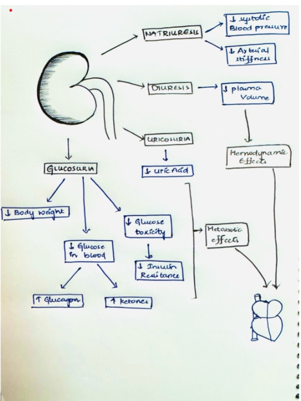
Beyond glycemic control, SGLT2 inhibitors like dapagliflozin may exert beneficial effects in HF through mechanisms including improved cardiac remodeling, reduced sodium retention, decrease plasma volume, and potential metabolic modulation. These mechanisms contribute to improved hemodynamics and reduced cardiovascular morbidity and mortality, as observed in clinical trials.
Contraindication
eGFR < 30mL/minute/1.73 m2
Safety and Tolerability
Safety profiles across trials indicate that dapagliflozin is generally well tolerated in HF patients, with adverse event rates comparable to placebo. Common side effects include volume depletion and renal dysfunction (Acute kidney injury due to dehydration), which are manageable with appropriate monitoring. Other severe side effects are euglycemic diabetic ketoacidosis, allergic reaction and genital infections (mostly fungal).
Canagliflozin increase the risk of lower limb amputation and bone fractures.
Clinical Implications and Future Directions
The efficacy of SGLT2 inhibitors in reducing HF-related events underscores their potential as adjunctive therapy in standard HF management. Future research should focus on optimizing patient selection, exploring combination therapies, and elucidating long-term benefits and risks in diverse patient populations.
Conclusion
SGLT2 inhibitors like dapagliflozin show great promise in improving outcomes for patients with heart failure. These medications not only reduce cardiovascular risks but also improve quality of life, highlighting their potential as essential elements of comprehensive heart failure treatment strategies.
References
McMurray JJV, Solomon SD, Inzucchi SE, Køber L, Kosiborod MN, Martinez FA, Ponikowski P, Sabatine MS, Anand IS, Bělohlávek J, Böhm M, Chiang CE, Chopra VK, de Boer RA, Desai AS, Diez M, Drozdz J, Dukát A, Ge J, Howlett JG, Katova T, Kitakaze M, Ljungman CEA, Merkely B, Nicolau JC, O’Meara E, Petrie MC, Vinh PN, Schou M, Tereshchenko S, Verma S, Held C, DeMets DL, Docherty KF, Jhund PS, Bengtsson O, Sjöstrand M, Langkilde AM; DAPA-HF Trial Committees and Investigators. Dapagliflozin in Patients with Heart Failure and Reduced Ejection Fraction. N Engl J Med. 2019 Nov 21;381(21):1995-2008. doi: 10.1056/NEJMoa1911303. Epub 2019 Sep 19. PMID: 31535829.
Solomon SD, McMurray JJV, Claggett B, de Boer RA, DeMets D, Hernandez AF, Inzucchi SE, Kosiborod MN, Lam CSP, Martinez F, Shah SJ, Desai AS, Jhund PS, Belohlavek J, Chiang CE, Borleffs CJW, Comin-Colet J, Dobreanu D, Drozdz J, Fang JC, Alcocer-Gamba MA, Al Habeeb W, Han Y, Cabrera Honorio JW, Janssens SP, Katova T, Kitakaze M, Merkely B, O’Meara E, Saraiva JFK, Tereshchenko SN, Thierer J, Vaduganathan M, Vardeny O, Verma S, Pham VN, Wilderäng U, Zaozerska N, Bachus E, Lindholm D, Petersson M, Langkilde AM; DELIVER Trial Committees and Investigators. Dapagliflozin in Heart Failure with Mildly Reduced or Preserved Ejection Fraction. N Engl J Med. 2022 Sep 22;387(12):1089-1098. doi: 10.1056/NEJMoa2206286. Epub 2022 Aug 27. PMID: 36027570.
Nassif ME, Windsor SL, Borlaug BA, Kitzman DW, Shah SJ, Tang F, Khariton Y, Malik AO, Khumri T, Umpierrez G, Lamba S, Sharma K, Khan SS, Chandra L, Gordon RA, Ryan JJ, Chaudhry SP, Joseph SM, Chow CH, Kanwar MK, Pursley M, Siraj ES, Lewis GD, Clemson BS, Fong M, Kosiborod MN. The SGLT2 inhibitor dapagliflozin in heart failure with preserved ejection fraction: a multicenter randomized trial. Nat Med. 2021 Nov;27(11):1954-1960. doi: 10.1038/s41591-021-01536-x. Epub 2021 Oct 28. PMID: 34711976; PMCID: PMC8604725.
Petrie MC, Verma S, Docherty KF, Inzucchi SE, Anand I, Belohlávek J, Böhm M, Chiang CE, Chopra VK, de Boer RA, Desai AS, Diez M, Drozdz J, Dukát A, Ge J, Howlett J, Katova T, Kitakaze M, Ljungman CEA, Merkely B, Nicolau JC, O’Meara E, Vinh PN, Schou M, Tereshchenko S, Køber L, Kosiborod MN, Langkilde AM, Martinez FA, Ponikowski P, Sabatine MS, Sjöstrand M, Solomon SD, Johanson P, Greasley PJ, Boulton D, Bengtsson O, Jhund PS, McMurray JJV. Effect of Dapagliflozin on Worsening Heart Failure and Cardiovascular Death in Patients With Heart Failure With and Without Diabetes. JAMA. 2020 Apr 14;323(14):1353-1368. doi: 10.1001/jama.2020.1906. Erratum in: JAMA. 2021 Apr 6;325(13):1335. doi: 10.1001/jama.2021.2802. PMID: 32219386; PMCID: PMC7157181.
Wiviott SD, Raz I, Bonaca MP, Mosenzon O, Kato ET, Cahn A, Silverman MG, Zelniker TA, Kuder JF, Murphy SA, Bhatt DL, Leiter LA, McGuire DK, Wilding JPH, Ruff CT, Gause-Nilsson IAM, Fredriksson M, Johansson PA, Langkilde AM, Sabatine MS; DECLARE–TIMI 58 Investigators. Dapagliflozin and Cardiovascular Outcomes in Type 2 Diabetes. N Engl J Med. 2019 Jan 24;380(4):347-357. doi: 10.1056/NEJMoa1812389. Epub 2018 Nov 10. PMID: 30415602.
Hema Manvi Koneru, MBBS Rajiv Gandhi Institute of Medical Sciences, Telangana, India.
Amanpreet Kaur, MBBS Government Medical College Patiala, India.
Divyasri Koneru, MBBS Dr.Pinnamaneni Siddhartha Institute of Medical Sciences and Research Foundation, India
Amin H. Karim MD FACC, Clinical assistant professor, Baylor College of Medicine, Houston, Texas.
A 74-year-old male presented with complaints of dizziness. His medical history includes hypertension, diabetes mellitus, mitral regurgitation, tricuspid regurgitation, and a transient ischemic attack five months prior. Additionally, he reported two episodes of memory lapses within the past year.
The patient denied experiencing orthopnea, paroxysmal nocturnal dyspnea, chest pain, smoking, shortness of breath, leg swelling, speech disturbances, disequilibrium, blurry vision, syncope, tinnitus, hearing loss, ataxia, numbness, tingling, pins, and needles in the arms or legs.
MEDICATIONS
Aspirin 81mg sustained-release oral tablet, once daily; atorvastatin calcium 80mg tablet, taken orally once daily; clopidogrel 75mg tablet, taken orally daily; dulaglutide 0.75mg/0.5ml subcutaneous pen injector, administered subcutaneously once weekly; gabapentin 300mg oral capsule, taken once daily; losartan 25mg tablet, taken orally once daily; metformin hydrochloride 750mg extended-release tablet, taken twice daily with meals; nitroglycerin 0.4 mg sublingual tablet, to be used when chest pain persists; and thiamine 100 mg oral tablet, taken once daily. These medications have been prescribed to effectively manage the patient’s medical conditions and symptoms.
The patient had a history of allergy to the contrast medium used for the radiological examination.
LAB WORKUP
HBA1C-6.6gm/dl
The rest of the blood reports- are within in normal range.
ELECTROCARDIOGRAM
Sinus tachycardia with nonspecific ST-abnormality
CTA CORONARY ARTERIES
There is severe coronary calcification. The observed calcium score is 780, which is at the 75th percentile for subjects of the same age, gender, race/ethnicity who are free of clinical cardiovascular disease and treated diabetes.
Moderate to severe, predominantly calcified. Coronary artery disease involving proximal and LAD with moderate luminal stenosis. FFR-CT was performed on the LAD which showed significant flow limitation of the mid-LAD (left anterior descending artery).
CTA OF BRAIN AND NECK
Brain–moderate chronic microvascular ischemia, suprasellar 8 x 7 mm partially calcified lipoma or dermoid cyst.
Neck-Complete occlusion of the left vertebral artery V1 and V2 segments. Atherosclerosis of bilateral extracranial carotid arteries without significant stenosis.
The head shows no significant stenosis or occlusion.
MRI BRAIN
No acute ischemia.
Hypothalamus showing non-enhancing mass suggestive of Lipoma/Dermoid cyst.
Moderate chronic microvascular ischemia.
ECHOCARDIOGRAM
Mild left ventricular hypertrophy.
Trace tricuspid regurgitation.
Mitral regurgitation.
Left ventricular dysfunction.
NUCLEAR STRESS TEST
A small area of perfusion defect in the inferior wall which fills with reperfusion
CAROTID ULTRASOUND
No occlusive disease.
Following a thorough workup and assessment of the patient’s clinical presentation, imaging studies, and diagnostic tests, a final diagnosis of subclavian steal syndrome was made. This condition is attributed to the underlying pathology of atherosclerosis which mainly affects the subclavian artery.
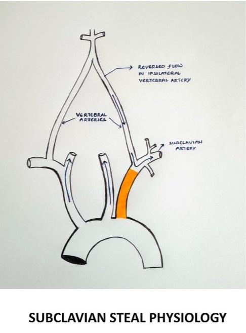
DEFINITION
It is characterized by the retrograde flow of blood in the vertebral artery, primarily resulting from significant stenosis or occlusion in the pre-vertebral subclavian artery resulting in cerebrovascular symptoms on the ipsilateral side of the occlusion.
§ During exertion, the subclavian artery steals blood from the vertebrobasilar artery circulation to supply the arm, which leads to vertebrobasilar insufficiency.
SUBCLAVIAN STEAL PHYSIOLOGY
EPIDEMIOLOGY
The prevalence of subclavian steal syndrome has not been well defined, although subclavian steal physiology has been reported in 1.3 to 2.6% of the general population of patients with extracranial atherosclerosis (1,2) as well as within subsets of those presenting with acute ischemic stroke (3)
ETIOLOGY
The most common cause is atherosclerosis.
Other vascular causes- Takayasu arteritis, giant cell arteritis
Arterial thoracic outlet syndrome
Congenital anomalies- anomalies of the aortic arch, anomalies of the brachiocephalic trunk
A consequence of corrective surgery- surgical repair of the Tetralogy of Fallot with a Blalock-Taussig anastomosis, surgical management of the coarctation of the aorta.
Subclavian stenosis more commonly occurs on the left side (>75 percent), possibly due to more acute origin on the left subclavian artery resulting in accelerated atherosclerosis from increased turbulence. (4,5,6)
CLINICAL FEATURES
Neurovascular symptoms can be caused by vertebrobasilar ischemia of the brainstem or cerebellum
Symptoms include dizziness, vertigo, binocular double vision, dysarthria, syncope, and drop attacks (sudden falls without loss of consciousness) (7)
Episodes can be precipitated by exercise of the ischemic arm and precipitated by certain head movements.
Atypical neurological presentations- there is also increasing recognition that patients with vertebrobasilar insufficiency can present with other types of symptoms not directly attributable to brainstem or cerebellar ischemia.
Subarachnoid hemorrhage (8)
Cognitive and mood abnormalities
EVALUATION
Although neurovascular symptoms associated with subclavian steal physiology occur in only a minority of patients, symptoms of vertebrobasilar ischemia demand critical evaluation (10,11)
When to suspect subclavian steal syndrome
Diagnosis should be considered in patients with a measurable upper extremity blood pressure differential who develop episodic neurological symptoms attributable to the brainstem or cerebellar ischemia.
Suspicion is increased if the episodes are provoked by arm exercise, especially if accompanied by ischemic arm symptoms (7)
PHYSICAL EXAMINATION
All major pulses should be palpated, and blood pressure should be checked in both arms.
It is important to examine the subclavian arteries in the supraclavicular fossa using palpation and auscultation for paraventricular bruits, also vertebral and carotid arteries for the evidence of any occlusive arterial disease.
On examination, there may be a significant decrease in the blood pressure on the affected side with a pulse delay appreciated when palpating the radial arteries simultaneously known as RADIO-RADIAL DELAY.
Check for any evidence of thromboembolism in the skin of the hands and nailbeds of the affected extremities.
Ischemia affecting the temporo-occipital areas of the cerebral hemispheres or segments of the brainstem and cerebellum characteristically produces bilateral symptoms.
RADIOLOGICAL EXAMINATION
Initial vascular imaging
1. Duplex ultrasound
A subclavian artery peak systolic velocity >240cm/sec is predictive of a significant (>70 percent) subclavian artery stenosis (9)
When severe stenosis (>80 percent narrowing) of the proximal subclavian artery is present, 65 percent of pa>ents have permanent flow reversal in the ipsilateral vertebral artery, and 30 percent have intermittent flow reversal (10,11)
2. Transcranial doppler ultrasound
Confirmatory vascular imaging
Computed tomographic angiography (CTA)-provides precise measurement of the severity of the stenosis. main drawback is the absence of dynamic flow information.
Magnetic resonance angiography (MRA)- sensitive than CTA. Does not depict images of arterial anatomy but rather the behavior and speed of the flowing protons in the vessel. Also provides information on the intracranial cerebrovascular circulation.
Catheter-based digital subtraction angiography (DSA)
DIAGNOSIS
DIAGNOSTIC CRITERIA– the presence of subclavian steal physiology with demonstration of a subclavian artery stenosis/ occlusion proximal to the origin of the vertebral artery causing
Marked reduction in the ipsilateral brachial artery blood pressure.
Reversal of the direction of blood flow in the ipsilateral vertebral artery
Neurological symptoms referrable to the vertebrobasilar circulation (cerebellum, brainstem, thalamus, occipital regions)
The likelihood that the presence of subclavian steal physiology will lead to the subclavian steal syndrome increases as the brachial pressure differential between the two limbs becomes more pronounced, particularly >40mmhg (5)
DIFFERENTIAL DIAGNOSIS
These include all the potential causes of vertebrobasilar embolism due to atherosclerosis, hypertension, hypercoagulable states, tumors, tobacco smoking, trauma, and others
MANAGEMENT
Management of patients with subclavian steal syndrome is individualized depending on the etiology, type, and severity of symptoms and their impact on quality of life
ATHEROSCLEROTIC SUBCLAVIAN STEAL SYNDROMEAPPROACH
For most patients with atherosclerosis as the etiology for subclavian steal syndrome, conservative management is the preferred initial therapy.
Initial medical management
Start with the low-dose aspirin.
However, the addition of other anti-platelet drugs does not seem justified since this condition is hemodynamic derangement. once a decision is made for surgical management, additional anti-thrombotic agents are required.
Optimal blood pressure management is required within the ipsilateral side brachial artery.
Surgical management
Proximal subclavian endarterectomy, which is a trans-thoracic approach used for revascularizing the subclavian artery.
Re-vascularization surgery via endovascular techniques (carotid-subclavian bypass or subclavian-carotid transposition).
Carotid intervention in patients with severe concomitant carotid artery disease helps to improve cerebral perfusion.
NON-ATHEROSCLEROTIC SUBCLAVIAN STEAL SYNDROME
For non-atherosclerotic subclavian syndrome etiologies, the primary cause needs to be addressed.
Example- Takayasu arteritis is by systemic glucocorticoids or steroid-sparing agents like biological DMARD (Disease Modifying Anti-Rheumatoid Drugs) or non-biological DMARD
CONCLUSION
This case highlights the importance of a detailed evaluation of subclavian steal syndrome can be due to atherosclerosis or non-atherosclerotic causes like Takayasu arteritis, giant cell arteritis, arterial thoracic outlet syndrome, and other causes. In our case, it might be a subclavian steal syndrome secondary to atherosclerosis.
REFERENCES
Fields WS, Lemak NA. Joint Study of extracranial arterial occlusion. VII. Subclavian steal–a review of 168 cases. JAMA. 1972 Nov 27;222(9):1139-43. PMID: 4678043.
Hennerici M, Klemm C, Rautenberg W. The subclavian steal phenomenon: a common vascular disorder with rare neurologic deficits. Neurology. 1988 May;38(5):669-73. doi: 10.1212/wnl.38.5.669. PMID: 3362359.
Bajko Z, Motataianu A, Stoian A, Barcutean L, Andone S, Maier S, Drăghici IA, Cioban A, Balasa R. Prevalence and Clinical Characteristics of Subclavian Steal Phenomenon/Syndrome in Patients with Acute Ischemic Stroke. J Clin Med. 2021 Nov 10;10(22):5237. doi: 10.3390/jcm10225237. PMID: 34830519; PMCID: PMC8621575.
Ochoa VM, Yeghiazarians Y. Subclavian artery stenosis: a review for the vascular medicine practitioner. Vasc Med. 2011 Feb;16(1):29-34. doi: 10.1177/1358863X10384174. Epub 2010 Nov 15. PMID: 21078767.
Labropoulos N, Nandivada P, Bekelis K. Prevalence and impact of the subclavian steal syndrome. Ann Surg. 2010 Jul;252(1):166-70. doi: 10.1097/SLA.0b013e3181e3375a. PMID: 20531004.
Shadman R, Criqui MH, Bundens WP, Fronek A, Denenberg JO, Gamst AC, McDermott MM. Subclavian artery stenosis: prevalence, risk factors, and association with cardiovascular diseases. J Am Coll Cardiol. 2004 Aug 4;44(3):618-23. doi: 10.1016/j.jacc.2004.04.044. PMID: 15358030.
KESTELOOT H, VANHOUTE O. REVERSED CIRCULATION THROUGH THE VERTEBRAL ARTERY. Acta Cardiol. 1963;18:285-99. PMID: 14045892.
Rodriguez-Lopez JA, Werner A, Martinez R, Torruella LJ, Ray LI, Diethrich EB. Stenting for atherosclerotic occlusive disease of the subclavian artery. Ann Vasc Surg. 1999 May;13(3):254-60. doi: 10.1007/s100169900254. PMID: 10347257.
Gutierrez GR, Mahrer P, Aharonian V, Mansukhani P, Bruss J. Prevalence of subclavian artery stenosis in patients with peripheral vascular disease. Angiology. 2001 Mar;52(3):189-94. doi: 10.1177/000331970105200305. PMID: 11269782.
Osiro S, Zurada A, Gielecki J, Shoja MM, Tubbs RS, Loukas M. A review of subclavian steal syndrome with clinical correlation. Med Sci Monit. 2012 May;18(5):RA57-63. doi: 10.12659/msm.882721. PMID: 22534720; PMCID: PMC3560638.
Cornelissen SA, Heye S, Maleux G, Daenens K, van Loon J, De Vleeschouwer S. Treatment of ruptured subclavian steal flow-related vertebrobasilar junction aneurysms: Case report on surgical and endovascular considerations from two cases. Int J Surg Case Rep. 2022 Jan;90:106744. doi: 10.1016/j.ijscr.2021.106744. Epub 2021 Dec 30. PMID: 34991048; PMCID: PMC8741505.
Ahuja CK, Joshi M, Mohindra S, Khandelwal N. Vertebrobasilar Junction Aneurysm Associated with Subclavian Steal: Yet another Hemodynamic Cause for Aneurysm Development and Associated Challenges. Neurol India. 2020 May-Jun;68(3):708-709. doi: 10.4103/0028-3886.288981. PMID: 32643699.
Tonetti DA, Jankowitz BT. Subclavian Steal Flow-Related Aneurysm Formation. World Neurosurg. 2019 May;125:101-103. doi: 10.1016/j.wneu.2019.01.186. Epub 2019 Feb 8. PMID: 30743034.
Mousa AY, Morkous R, Broce M, Yacoub M, Sticco A, Viradia R, Bates MC, AbuRahma AF. Validation of subclavian duplex velocity criteria to grade severity of subclavian artery stenosis. J Vasc Surg. 2017 Jun;65(6):1779-1785. doi: 10.1016/j.jvs.2016.12.098. Epub 2017 Feb 17. PMID: 28222983.
Nicholls SC, Koutlas TC, Strandness DE. Clinical significance of retrograde flow in the vertebral artery. Ann Vasc Surg. 1991 Jul;5(4):331-6. doi: 10.1007/BF02015293. PMID: 1878290.
Harper C, Cardullo PA, Weyman AK, Patterson RB. Transcranial Doppler ultrasonography of the basilar artery in patients with retrograde vertebral artery flow. J Vasc Surg. 2008 Oct;48(4):859-64. doi: 10.1016/j.jvs.2008.05.057. Epub 2008 Aug 9. PMID: 18692344.
From Accidents to Advancements in the Realm of Medications.
Mahmood Syed, MS4 American University School of Medicine
Amanpreet Kaur, MBBS Government Medical College Patiala, India
Amin H. Karim MD FACC Clinical Assistant Professor Baylor College of Medicine, Houston, Texas.
“When you have acquired knowledge and experience, it is very pleasant to break the rules and to be able to find something nobody has thought of.” These were the words of renowned Bacteriologist, Andrew Fleming, who discovered Penicillin widely used today. There are drugs whose original discovery was intended for specific indications. However, their side effects were so useful that these medications were eventually repurposed as treatments for other medical conditions. This phenomenon is also known as “drug repurposing” or “serendipitous discovery”. Here are some examples of such medications widely used today:
The discovery of Minoxidil is a fascinating chapter in medicinal advancements. It was initially created for the treatment of Hypertension. However, during clinical studies, scientists discovered a surprising adverse effect that caused hair growth in patients that were taking this medication. This coincidental finding prompted more research into Minoxidil’s potential as a male pattern baldness remedy. Eventually, it was repurposed for this indication.
Viagra (Sildenafil) is another interesting medication that was unintentionally found while being used in the treatment of Angina Pectoris and Pulmonary Hypertension. An unanticipated side effect that the researchers discovered during clinical trials was that many of the male participants reported having better erectile function after taking this medication due to its vasodilatory effects. Therefore, it was repurposed as a medication for men suffering from Erectile Dysfunction (ED).
Bimatoprost was another medication that was initially discovered for the treatment of Glaucoma, but researchers noted that it increased eyelash length, thickness, and blackness in the patients who received these eye drops for the treatment of Glaucoma. This led to its use for cosmetic enhancement of eyelashes today.
Ozempic (Semaglutide) was originally discovered for the treatment of Type 2 Diabetes Mellitus. This medication functioned as a GLP-1 agonist. Researchers found that it caused significant weight reduction in addition to controlling blood glucose levels. After further clinical trials, it was approved as a medication for weight loss.
Gabapentin was initially discovered as a potential treatment for seizures as it was structurally similar to the neurotransmitter GABA. After its initial discovery, Gabapentin was subsequently found to have efficacy in treating neuropathic pain in patients who were treated for seizures, leading to its approval for this indication as well.
Finasteride was initially investigated for its potential use in the treatment of Benign Prostatic Hyperplasia. While clinical trials were ongoing to see the effectiveness of this medication for BPH, it was observed that patients taking finasteride experienced hair growth as a side effect. Eventually, it was repurposed for the treatment of hair loss.
Methotrexate, a potent inhibitor of Folate metabolism, was used for the treatment of many cancers including Acute Lymphocytic Leukemia (ALL) and Choriocarcinoma. It was discovered to be effective in treating autoimmune conditions due to its ability to inhibit cell growth and modulate immune responses. Today, it is used to treat conditions like Systemic Lupus Erythematosus (SLE) and Rheumatoid Arthritis (RA).
The development of aspirin from a painkiller to a multi-indicated drug is evidence regarding its healing characteristics. Initially, aspirin was found to be useful for reducing fever and pain. However, it is now essential for maintaining cardiovascular health, controlling inflammation, and even preventing and reducing the incidence of cancer, particularly in the setting of colon cancer. This extensive range of applications highlights the significance of aspirin in modern medicine and its ongoing importance over a century after it was initially discovered.
There are other medications that produce treatment indications as a result of their side effect. In the world of ever-evolving medicine and advancements in medical research, there is no doubt there will be more discoveries of newer medications as well as current medications with newer indications. The medical field is expanding on a level never experienced before in humanity. As new therapies become available, as well as more research being generated as a result of clinical trials and controlled studies, we will see an even greater advancement in medicine in the near future than we have ever seen before. The only limitation that we have is the limitations we set on ourselves. In the book, The Laws of Medicine, by Pulitzer Prize-winning author Siddhartha Mukherjee, he says “ In medicine, mistakes are inevitable but learning from them is essential.” Furthermore, if these mistakes lead to newer discoveries and treatments in the realm of medicine, then mistakes might just be better than getting it right the first time.